
CT Scans
@diagnosis_cirrhosis “I’m so sorry…wait, cirrhosis?” 🤦🏻♀️😡 I had a CT scan today. Afterward, I was sitting in the little observation area where they keep you for a few minutes after contrast. Someone overheard me talking about my hair falling out. She assumed I had cancer. Immediately, her face softened. “I’m so sorry.” She asked questions. She was kind. She was genuinely empathetic. Then she asked what kind of cancer it was. I told her I didn’t have cancer. I have cirrhosis. And I watched the temperature in the room change. Not dramatically. Just enough. Enough that if you’ve lived with liver disease, you know exactly what I mean. — I think one of the hardest parts of cirrhosis isn’t always the disease. Sometimes it’s the story people write about you before you’ve said a single word. If you’ve ever wondered: 👉 Why does society stigmatize alcoholism? 👉 Why is there so much stigma around liver disease? 👉 Why do people think liver disease is self imposed? 👉 Why does liver disease seem to get less empathy than other illnesses? You’re not imagining it. Those conversations happen more often than people realize. — She was trying to course correct, She said, “My cousin had cirrhosis…he was an alcoholic.” There was almost an apology built into the sentence. As though she was trying to figure out which category to place me in. And all I could think to say was, “I wish I could have met him. He sounds like a nice guy.” Because he probably was. — One of the biggest misconceptions about cirrhosis is that it’s one disease with one cause. It isn’t. Cirrhosis is the final stage of scarring from many different liver diseases. Autoimmune hepatitis. Fatty liver disease. Hepatitis B. Hepatitis C. Primary biliary cholangitis. Primary sclerosing cholangitis. Genetic diseases like hemochromatosis or Wilson’s disease. Alcohol-related liver disease. Drug-induced liver injury. And many others. Different roads. Same destination. — And even if someone’s cirrhosis is related to alcohol… Why would that make them less deserving of compassion? Alcohol use disorder is a medical condition. Recovery is hard. Living with cirrhosis is hard. Those things deserve empathy too. I’ve never been offended when someone assumes alcohol caused my cirrhosis. Not because they’re correct. Because I don’t think people living with alcohol use disorder deserve less kindness than I do. They don’t. — Over time, these conversations have become easier. Not because the stigma disappeared. Because I stopped feeling responsible for carrying it. I’ve realized I don’t have to defend my diagnosis. I don’t have to earn compassion by explaining my medical history. Illness isn’t a morality test. And empathy shouldn’t depend on how someone became sick. — I think that’s what stayed with me after today’s scan. Not that someone made an assumption. We all do. It was realizing how quickly empathy and sympathy can become conditional. How easily our brains sort people into categories of deserving and undeserving. Cancer. Cirrhosis. Alcohol. Autoimmune. As though suffering becomes easier to measure once we’ve decided who is responsible. But illness doesn’t work that way. Neither should compassion. — 🧬 Common causes of cirrhosis include autoimmune hepatitis 🔥 fatty liver disease / MASLD / NAFLD / MASH / NASH 🍩 alcohol-related liver disease 🚫🍷 hepatitis B 🦠 hepatitis C 🦠 primary biliary cholangitis (PBC) 🧬 primary sclerosing cholangitis (PSC) 🧬 hemochromatosis ⚙️ Wilson’s disease 🪙 alpha-1 antitrypsin deficiency 🧩 drug-induced liver injury 💊 cryptogenic cirrhosis ❓ or other causes. 👩⚕️👨⚕️🩺 Always talk to your doctor or hepatologist about your liver disease, its cause, treatment options, and available support. And if you’re living with alcohol use disorder, know that you deserve compassionate medical care and empathy just as much as anyone else. Because the cause of an illness may shape the treatment. It should never determine the kindness someone receives. 💚 #liverhealth #alcoholism #cirrhosis #fattyliver #kindnessisfree ♬ Happy Time - Nazar
Top 4 Need to Knows
Important Medical Disclaimer: I am Angie, a cirrhosis patient sharing my personal lifestyle, nutrition, and mindset journey. I am a data aggregator and community builder, not a licensed medical professional, doctor, or dietitian. Content on diagnosiscirrhosis.com is based solely on my personal experience living with advanced liver disease and is intended for informational and educational purposes only. It does not constitute medical advice or a substitute for professional medical care. Always follow the recommendations of your own healthcare team and consult your hepatologist or physician before changing your diet, medication, or lifestyle routine.
My Experience with It
By Angie from @Diagnosis_Cirrhosis on TikTok
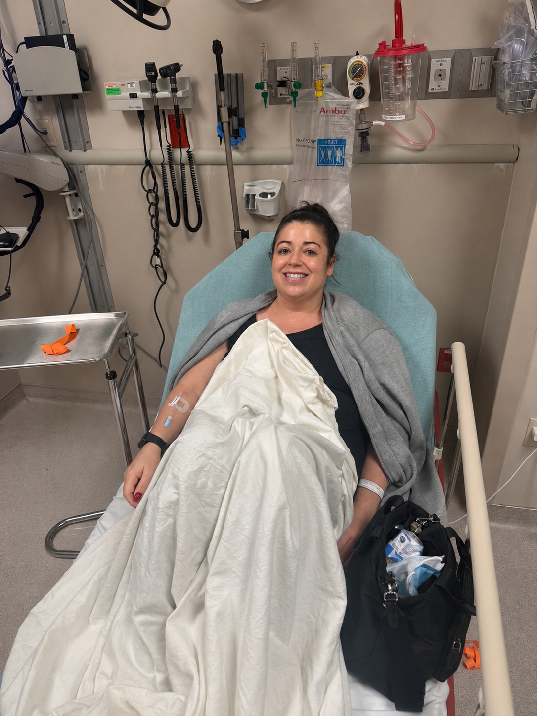
Here I am in Nov 2025, about a year after I decompensated in the ER for a CT to check for potential clots identified in an ultrasound
Before cirrhosis, CT was a guy from The Real World moreso than any medical fixture in my life.
Like, I knew it was imaging. I knew it was probably related to MRI in some vague way.
I could have told you it was a distant cousin of the MRI.
Then I got sick.
And once you get sick-sick, especially liver sick, especially portal hypertension /upper abdominal pain /what-is-my-body-doing-today sick, you start learning the emergency room protocol whether you wanted to enroll in that class or not.
Upper abdominal pain?
Right upper quadrant in question?
Liver area acting suspicious?
Welcome to the ER. Please enjoy your hospital bracelet.
Now please proceed to the picture machine.
And that is really how CT entered my life. Not as this huge, dramatic, scheduled event like my first MRI. CT felt more like part of the ER choreography. You go in. They ask questions. They poke. They press. They run bloodwork. They look concerned in that specific ER way where they are calm but somehow making you more nervous. And then eventually someone says, “We’re going to get a CT.”
And you are like, okay.
Apparently, we are getting shoved through the picture donut.
Because that is what it feels like.
It is not the MRI, the giant tube, the loud magnetic coffin. It’s the picture donut.
What It Feels Like
It is more like a little circle.
A hepatic investigative hoop. A bubble wand.
One of those old-fashioned bubble things you blow through, except instead of making bubbles, it is taking highly detailed pictures of your insides while you lie there wondering whether you are breathing deep enough or the same every time.
And honestly? CT is fast. That is the thing.
It is fast in a way MRI is absolutely not fast. MRI is an event. MRI has chapters. MRI has a soundtrack. MRI has character development.
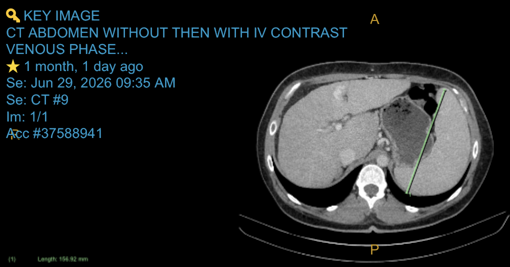
One example of a CT scan, this one featuring my enlarged spleen
CT is like:
Lay down.
Hold still.
Breathe in.
Hold it.
Contrast.
Warmth.
Maybe pee?
Done.
And yes, the contrast is weird. It can make you feel warm all over. It can give you that very specific, very rude sensation that you have just peed on the table.
But let’s be honest, I have to pee all the time anyway. Diuretics, hello.
The Vascular Veritas
It might be ordered by a transplant center because they want a sharper look at your blood vessels, shunts, portal vein flow, varices, anatomy, or whatever vascular chaos your body has decided to audition that season.
For me, I have shunts that get watched. So now CT is not just an ER thing.
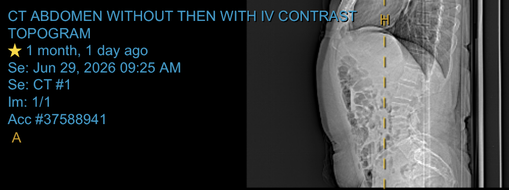
Some images make a lot less sense to stare at. What’s going on here? Still haven’t gotten to the bottom of the angle :)
It is your body building side roads because the main highway is a traffic disaster.
Portal hypertension is not subtle. It does not simply inconvenience the system. It starts rerouting blood like a deginerate city planner with a clipboard.
Compared to MRI
From the patient side, I want you to know this:.
It is quick.
It is usually painless.
It is not a tube in the MRI sense.
It is not 45 minutes of mechanical Berlin grunge.
And if you are looking back through your records trying to figure out, “Was that a CT or was that an MRI?” check the context.
Was it in the ER?
Was it fast?
Were they trying to rule something out quickly?
Was there abdominal pain, infection concern, bleeding concern, fluid concern, or some sudden hospital drama?
There is a decent chance that was CT.
Key Takeaways:
Find Out More
What the Procedure Is
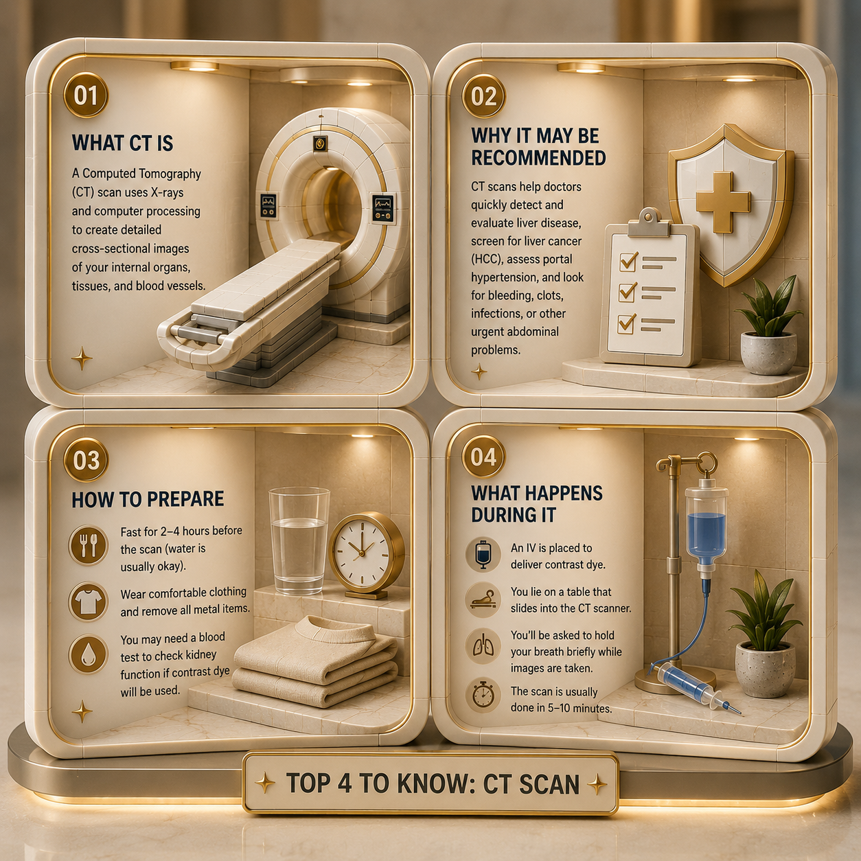
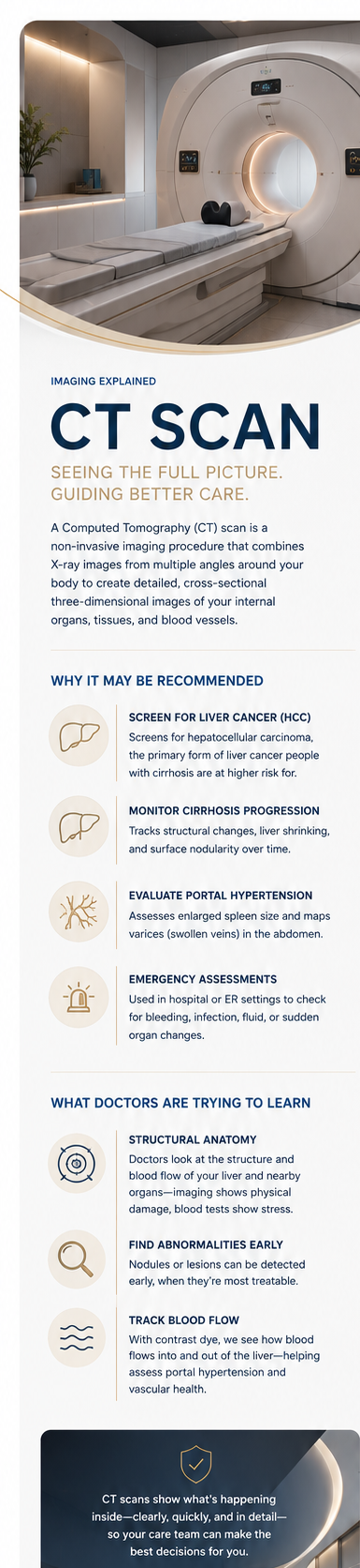
A Computed Tomography, or CT, scan is a non-invasive diagnostic imaging procedure that combines a series of X-ray images taken from multiple angles around your body [1]. Advanced computer processing merges these slices to create highly detailed, cross-sectional three-dimensional images of your internal soft tissues, organs, and blood vessels [2].
Why This Procedure May Be Recommended
In the management of advanced liver disease and cirrhosis, standard clinical protocols mandate regular abdominal imaging. A healthcare provider will typically recommend a CT scan to:
Screen for Hepatocellular Carcinoma, or HCC, which is the primary form of liver cancer that individuals with cirrhosis carry a higher risk of developing [3].
Monitor the Physical Progression of Cirrhosis, allowing doctors to evaluate structural changes, liver shrinking, or surface nodularity [4].
Evaluate Portal Hypertension complications, such as tracking the exact size of an enlarged spleen or mapping out dangerous varices, or swollen veins, in the abdomen [3].
Provide Emergency Assessments during Acute Hospital or ER Admissions to quickly check for internal bleeding, fluid infection, or sudden organ changes [5]
What Doctors Are Trying to Learn
Physicians use an abdominal CT scan to look directly at the structural anatomy and blood flow dynamics of your liver and surrounding organs [2]. Unlike blood tests that reveal cellular stress or manufacturing drops, imaging shows the physical damage [4].
Hepatologists are trying to identify any abnormal tissue nodules or lesions early, when they are small and highly treatable [3]. Furthermore, by using an injected contrast dye, doctors track the exact speed at which blood flows into the liver via the hepatic artery and drains through the portal vein [1]. This blood-flow mapping helps transplant centers evaluate vascular health and accurately judge the severity of portal hypertension [3].
How to Prepare
An abdominal CT scan requires specific preparation steps to ensure the internal images are sharp and clear:
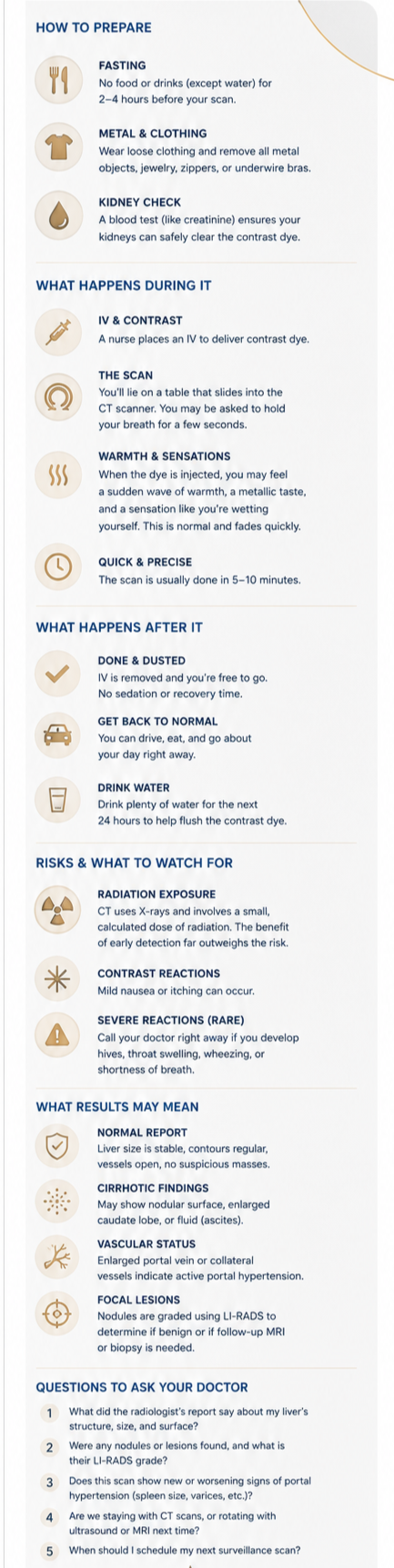
Fasting Requirements: You will generally be instructed to fast, meaning no food or liquids except water, for 2 to 4 hours prior to the scan to ensure your stomach and bowels are empty [5].
Metal and Clothing: Wear loose, comfortable clothing and be prepared to remove all metal objects, jewelry, zippers, or underwire bras, as metal severely distorts X-ray images [1].
Kidney Function Checks: Because contrast dye is cleared through your urinary system, your doctor will run a quick blood test, like creatinine, beforehand to ensure your kidneys are healthy enough to filter the fluid [2].
What Happens During and After
The procedure takes place in a dedicated imaging suite and is remarkably fast [5]. A nurse will place a standard intravenous, or IV, line in your arm to deliver the contrast dye [1]. You will lie flat on a motorized table that slides smoothly into the center of a large, doughnut-shaped CT scanner [5].
As the scanner rotates around you, the machine will play automated voice commands telling you exactly when to breathe in, hold your breath, and breathe normally to keep your organs from moving [2]. When the contrast dye is injected through your IV, you will feel a sudden, intense wave of warmth spread through your chest and body, often accompanied by a metallic taste in your mouth and a strange sensation that makes you feel like you are wetting yourself [1]. This feeling is completely harmless and fades in less than a minute. The active scanning is usually finished in 5 to 10 minutes.
Once the scan is complete, the table slides out, the IV line is removed, and you are free to sit up [5]. There is no recovery period or sedation involved, meaning you can drive yourself home immediately, eat a regular meal, and go about your day [2]. You will be advised to drink plenty of water over the next 24 hours to help your kidneys quickly flush the remaining contrast dye out of your system [1]. Be mindful of what “a lot” means if you have a fluid restriction.
Risks and What to Watch For
A CT scan is a highly controlled, safe clinical procedure [1]. Potential side effects and risks are minimal and include:
Radiation Exposure: CT scans use X-rays, meaning they expose you to a small, calculated dose of ionizing radiation [5]. In cirrhosis, the vital medical benefit of catching liver cancer early heavily outweighs this minimal risk [3].
Contrast Reactions: Some individuals may experience mild nausea or a localized itch from the dye [2].
Signs to watch for: Severe allergic reactions to contrast are very rare. However, contact your doctor immediately if you develop hives, swelling in your throat, wheezing, or shortness of breath after leaving the clinic [5].
What Results May Mean
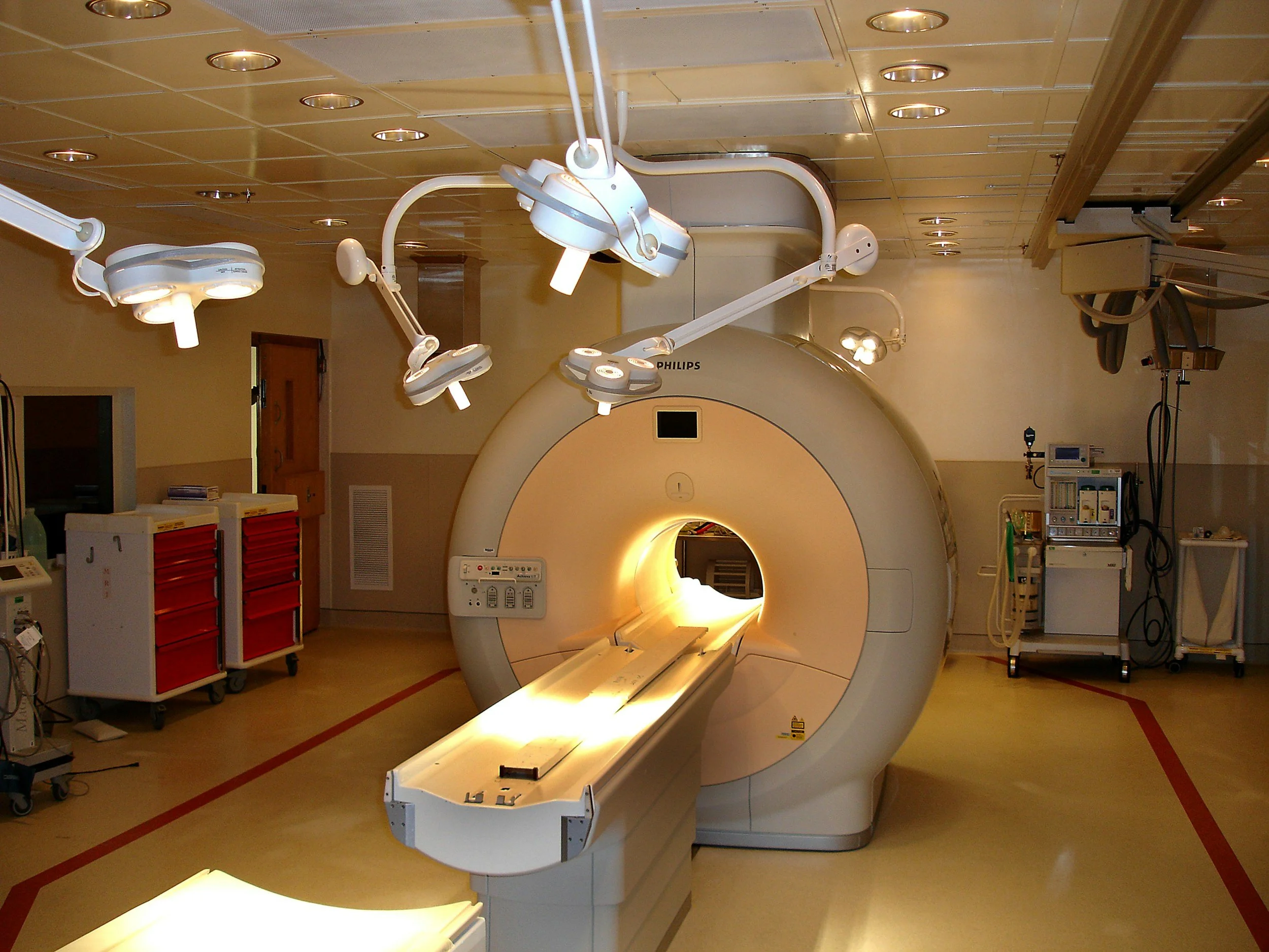
A specialized medical doctor called a radiologist will interpret the cross-sectional images and send a formal text report to your hepatologist [5].
A Normal Report
This indicates that your liver size is stable, the organ contours appear regular, blood vessels are open, and there are no suspicious focal masses or lesions detected [2].
Cirrhotic Structural Findings
The report may describe a “nodular or coarse liver surface,” an enlarged caudate lobe, or fluid accumulation, called ascites, which are all standard structural hallmarks of advanced liver disease [4].
Vascular Stating
Findings like an enlarged portal vein or prominent collateral vessels tell your care team that portal hypertension is active and requires continued medical management [3].
Focal Lesions
If a nodule is found, the radiologist will grade it using a standard system called LI-RADS, or Liver Imaging Reporting and Data System, to tell your doctor whether the spot is a benign scar or requires a follow-up MRI or biopsy [3].
Questions to Ask Your Doctor
To help build your personal tracking records and gain clarity at your next follow-up appointment, consider asking your doctor these questions:
What did the radiologist’s report state regarding the overall structure, size, and surface appearance of my liver today?
Were there any focal lesions or nodules detected on this scan, and if so, what was their specific LI-RADS grading?
Does this CT scan show any new or worsening signs of portal hypertension, such as an increase in spleen size or the development of abdominal varices?
Given my personal health plan, are we keeping my screening schedule strictly on CT scans, or are we rotating between ultrasound and MRI next time?
When is the optimal time for us to schedule my next routine surveillance scan to ensure we are staying completely ahead of any changes?
Sources
[1] : Read about contrast agents, the physical sensations they cause, and baseline imaging precautions via the Mayo Clinic CT Scan Guide.
[2]: Review the clinical expectations, cross-sectional formatting, and dye processing parameters on the Cleveland Clinic Computed Tomography Reference Portal. [1]
[3] Review the standardized rules for tracking observations under 10 mm and visualization criteria via the American College of Radiology US LI-RADS Update on RSNA
[4] Access the core repository for comprehensive guidelines regarding liver abnormalities, staging, and hepatic evaluations through the official American College of Gastroenterology (ACG) Guidelines Index.
[5] Study the baseline preparation instructions, cross-sectional belly scans, and fasting protocols at the National Institutes of Health MedlinePlus Abdominal CT Database.
