
Cirrhosis vs. Fibrosis

Important Medical Disclaimer: I am Angie, from @diagnosis_cirrhosis on TikTok. I am a cirrhosis patient sharing my personal lifestyle, nutrition, and mindset journey. I am a data aggregator and community builder, not a licensed medical professional, doctor, or dietitian. Content on diagnosiscirrhosis.com is based solely on my personal experience living with advanced liver disease and is intended for informational and educational purposes only. It does not constitute medical advice or a substitute for professional medical care. Always follow the recommendations of your own healthcare team and consult your hepatologist or physician before changing your diet, medication, or lifestyle routine.
The Difference of Reversibility
One day your routine lab work comes back slightly elevated. The next, you are looking at a medical report filled with staging terms like "bridging fibrosis" or "compensated cirrhosis," trying to figure out if your liver is facing a temporary hurdle or a permanent crisis. [1, 2]
@diagnosis_cirrhosis 🚨🧬 Fibrosis is NOT a life sentence… and it doesn’t automatically mean cirrhosis 👀 🎥 Live highlight 🎥 Hearing “liver damage” can feel like everything is already decided… but that’s not how this works 🚫 Fibrosis is the stage before cirrhosis — and in many cases, it can be slowed, stopped, or even reversed depending on the cause and how early it’s caught 📉📈 Progression is not guaranteed. Here’s what that actually means 👇 👉 Fatty liver can improve 🍩➡️🥗 👉 Inflammation can calm down 🔥➡️🧘♀️ 👉 Scar tissue can partially regress over time 🧬⬇️ 👉 The liver can regain function if the root cause is addressed 💪 But here’s the key: education matters. Because a lot of people hear “fibrosis” and think it’s already the worst-case scenario 😔 When in reality, it can be the turning point. Cirrhosis doesn’t happen overnight ⏳ It’s a progression — and that means there’s time to intervene. If you’re dealing with liver disease from: 👉 MASLD (fatty liver) 🍩➡️🩺 👉 MASH/NASH 🧪 👉 Alcohol-related liver disease 🚫🍷 👉 Hepatitis B 🦠 👉 Hepatitis C 🦠 👉 Autoimmune hepatitis 🛡️🔥 👉 Primary biliary cholangitis (PBC) 🧬 👉 Primary sclerosing cholangitis (PSC) 🧬 👉 Hemochromatosis ⚙️ 👉 Wilson’s disease 🪙 👉 Alpha-1 antitrypsin deficiency 🧩 👉 Drug-induced liver injury 💊 👉 Or anything else ❓ —know this: it is not too late to take action. Being told to change your lifestyle, your diet, your habits… it’s not a punishment ❌ It’s an opportunity to protect your liver before it gets worse 💛 Always talk to your doctor or hepatologist 👩⚕️👨⚕️🩺 and follow their guidance — but don’t assume the outcome is already written. #livehighlights #tiktoklive #fattyliver #fibrosis #liverhealth ♬ The Best Time - Infraction Music
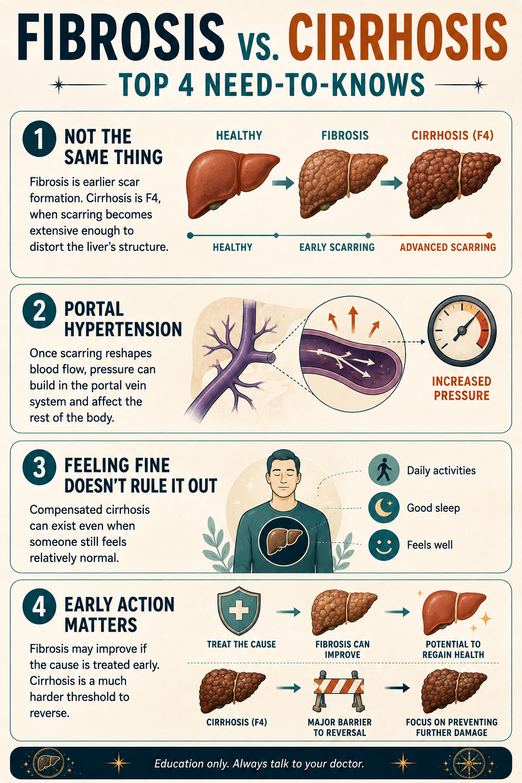
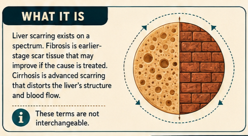
Liver scarring exists on a strict pathological spectrum.
Fibrosis is the initial, highly reversible accumulation of cellular scar tissue within the liver.
Cirrhosis is the advanced, often irreversible end-stage phase where widespread, structural scarring completely distorts the organ's anatomy,
as detailed by the NIH National Library of Medicine StatPearls Liver Cirrhosis Profile. [1, 2, 3]
People frequently use these terms interchangeably, but they represent two entirely different clinical realities. One is a quiet, manageable warning sign; the other is a profound mechanical failure that reshapes how your blood circulates and how your body processes toxins. [1, 2, 3]
What Is Liver Fibrosis
The liver possesses a remarkable, unique ability to regenerate. However, chronic injury—whether driven by metabolic dysfunction-associated steatotic liver disease (MASLD/NASH), prolonged alcohol use, viral hepatitis, autoimmune diseases or other causes—triggers a continuous cycle of inflammation. [1, 2, 3]
When liver cells (hepatocytes) are perpetually damaged, they release chemical signals that activate hepatic stellate cells.
As explained in a comprehensive MDPI Journal of Clinical Medicine Pathology Study, these activated cells transform into myofibroblast-like cells and begin pumping out excess extracellular matrix proteins, primarily collagen.
This is fibrosis—the microscopic laying down of scar tissue, akin to skin forming a hard scar after a deep cut. [1, 2, 3, 4]
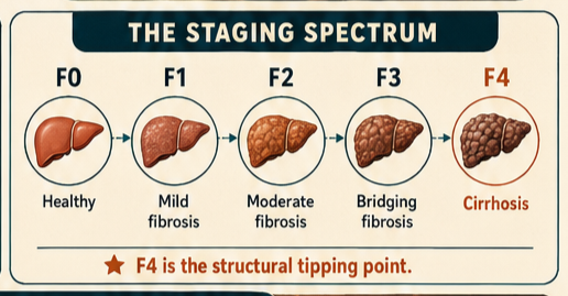
What are the Stages of Liver Scarring? (F0 to F4)
According to the historical METAVIR scoring system frequently utilized in clinical reviews, the progression of this tissue stiffness is tracked across a definitive medical gradient: [1]
Grade F0: No fibrosis; a healthy, fully compliant liver.
Grade F1: Portal fibrosis; mild scarring confined to the small blood vessels of the liver.
Grade F2: Few septa; moderate scarring that begins to extend out into the surrounding tissue.
Grade F3: Bridging fibrosis; advanced scarring where the bands of scar tissue interconnect, though the liver still maintains its basic shape.
Grade F4 (Cirrhosis): The structural tipping point. Widespread scarring has completely partitioned the liver into hard, bumpy "regenerative nodules," destroying its microscopic vascular layout. [1, 2, 3]
What Does Liver Fibrosis and Cirrhosis Feel Like?
The terrifying reality of early liver disease is its complete lack of physical feedback. Because the liver has no internal pain receptors, the transition through the early stages of scarring happens in total silence. [1]
During Fibrosis (Stages F1–F3), you frequently feel:
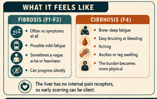
Absolutely nothing at all.
A vague, easily dismissed sense of mild, daily fatigue.
An occasional, dull ache or heavy sensation in the upper right side of your abdomen under your ribs. [1, 2]
When the disease crosses into Cirrhosis (Stage F4), patients experience:
A profound, bone-deep exhaustion that sleep cannot fix, driven by metabolic failure.
Sudden, unpredictable bruising or bleeding from minor bumps because the liver can no longer synthesize clotting proteins.
Severe, relentless skin itching (pruritus) caused by systemic bile salts backing up into the bloodstream.
Rapid, frightening physical changes like swelling in the ankles (edema) or a sudden, taut expansion of the abdomen (ascites). [1, 2, 3]
The Crossover Point: From Flexible Tissue to Mechanical Architecture Failure
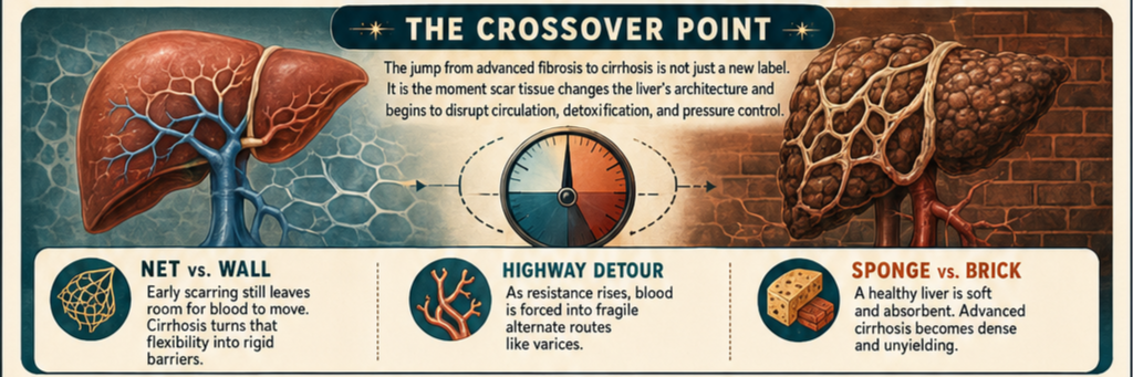
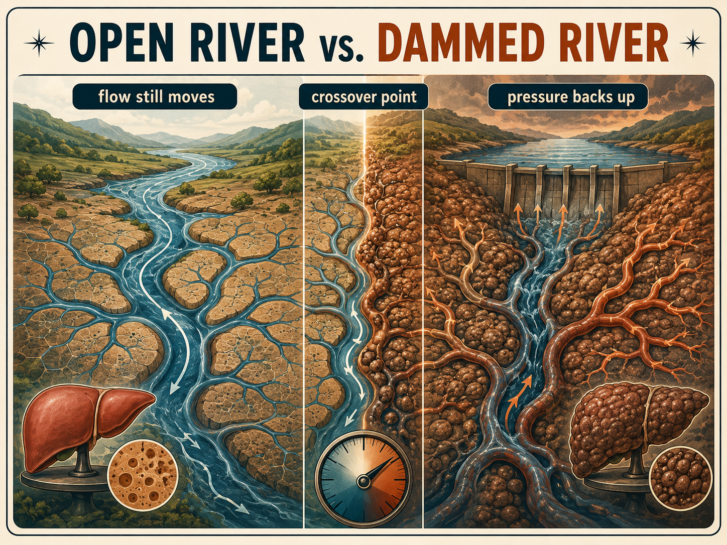
The transition from Stage F3 (advanced bridging fibrosis) to Stage F4 (cirrhosis) is not just a change in a letter grade; it is a profound structural and mechanical "crossover point" where the liver's fundamental anatomy collapses. [1, 2]
To visualize this crossover point, think of three concrete examples:
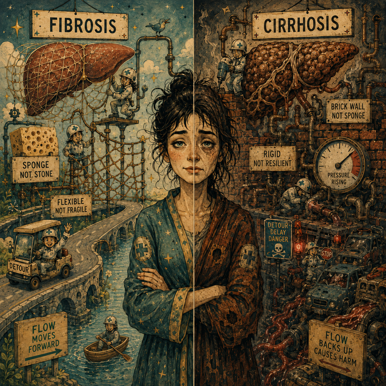
The Net vs. The Wall: In Stages F1 to F3, fibrosis is like a loose fishnet woven through the liver. Blood can still weave through the holes of the net, and healthy liver cells can breathe. At the F4 crossover point, the fishnet tightens, thickens, and turns into solid concrete walls. The blood can no longer flow around it, and the healthy tissue is permanently trapped in isolated pockets.
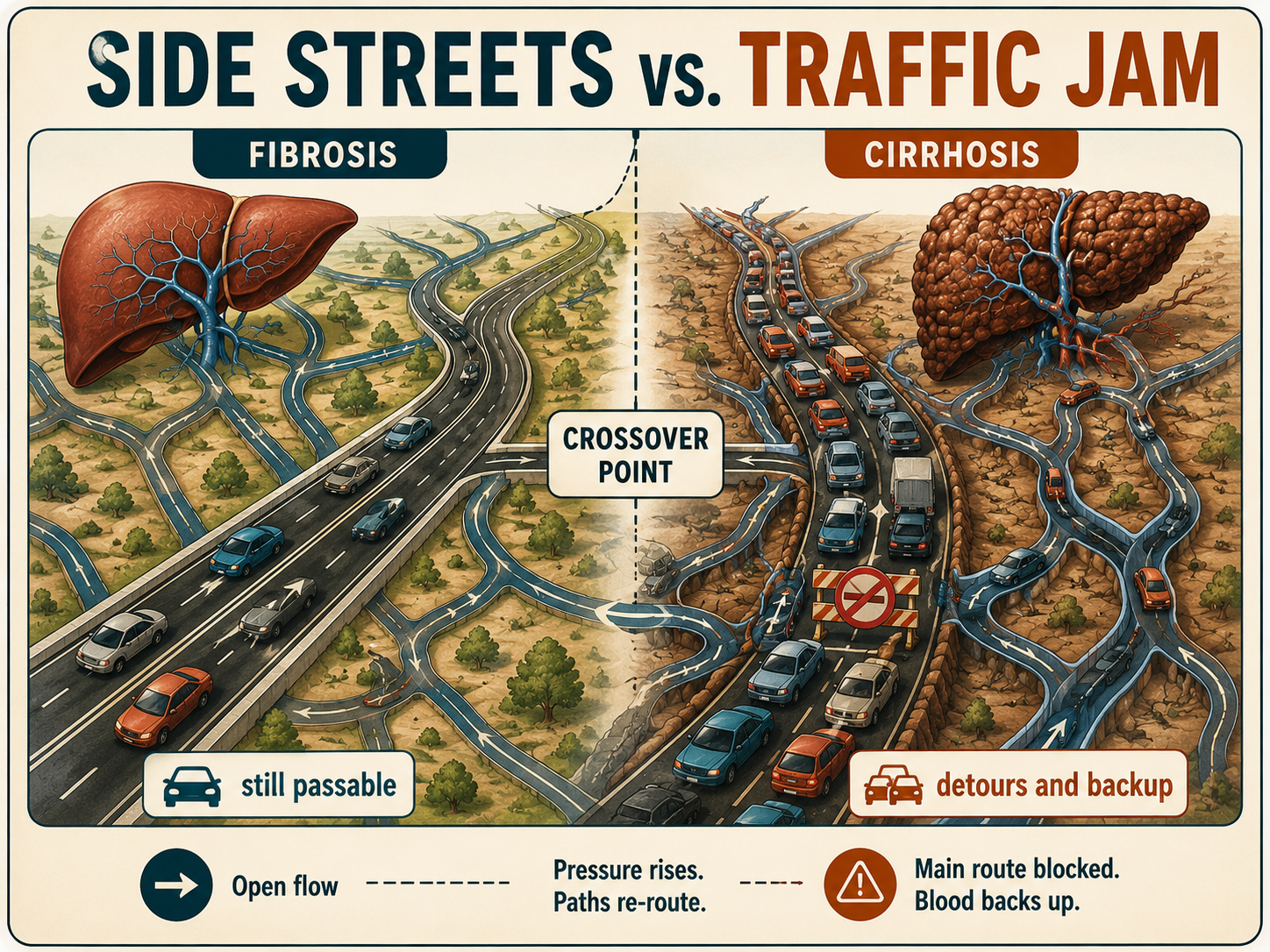
The Highway Detour: Imagine a multi-lane highway. In early fibrosis, a few lanes are closed for construction (scarring), causing mild traffic slows, but cars still reach their destination. At the cirrhosis crossover point, the entire highway is completely blocked off. Blood is forced to take volatile, unpaved side roads (esophageal varices) that were never built to handle high-speed traffic.
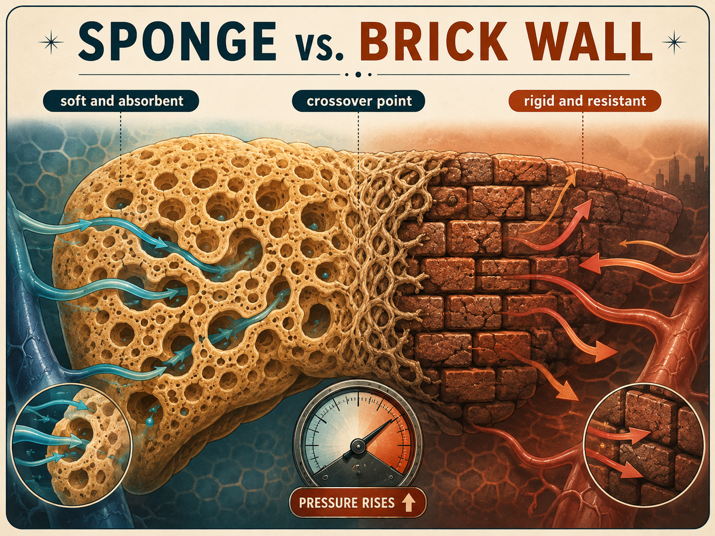
The Kitchen Sponge: A healthy liver is like a soft, damp kitchen sponge—flexible, compliant, and able to absorb blood effortlessly. In advanced fibrosis, parts of the sponge start to dry out and stiffen. At the cirrhosis crossover point, the sponge completely calcifies into a hard, dense brick. If you pour water over a brick, it cannot absorb it; the water simply reflects backward. [1, 2, 3, 4, 5, 6]
Clinical insights from an in-depth PMC Liver Fibrosis and Cirrhosis Regression Review note that before this crossover point, the liver's vascular matrix is elastic. Once the tissue crosses into true macronodular or micronodular cirrhosis, the sheer density of the collagen bands chokes off the liver's microcirculation, resulting in permanent structural distortion and a dangerous spike in internal blood pressure known as Clinically Significant Portal Hypertension (CSPH). [1, 2]
Things Nobody Explains about Liver Fibrosis
1. The "Compensated" Illusion
You can have full-blown stage F4 cirrhosis and feel completely healthy. This is known as compensated cirrhosis. The American Academy of Family Physicians Guide on Cirrhosis details how the remaining pockets of healthy liver tissue work overtime to keep up with your body's demands. However, it represents a fragile clinical equilibrium; a minor infection or medication stress can suddenly push the liver over the edge into decompensated cirrhosis, triggering acute liver failure. [1, 2]
2. Lab Blood Tests Can Be Misleading
Many patients assume that normal liver enzymes (AST and ALT) mean their liver is perfectly healthy. This is a dangerous misconception. As highlighted by the diagnostics reviewed in the PMC Pathology Study, active inflammation can quiet down as fibrosis progresses toward cirrhosis, causing enzyme levels to drop back into the "normal" clinical range. A liver can be severely scarred and cirrhotic even with pristine routine blood work.[1, 2, 3]
3. It Disrupts Your Entire Vascular Highway
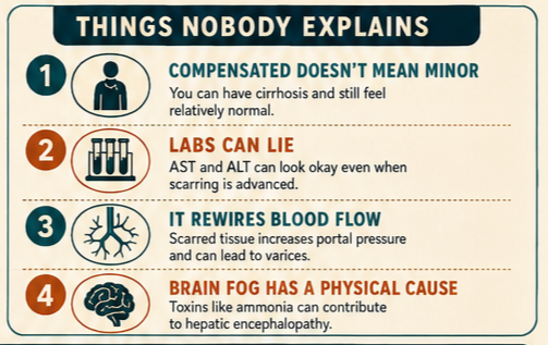
Cirrhosis is not just a localized liver problem; it is a cardiovascular crisis. Because blood cannot easily push through the hard, scarred liver tissue, it backs up into the portal vein. This high pressure forces the body to create fragile, bypass veins (varices) in the esophagus and stomach, which are highly susceptible to sudden, life-threatening ruptures. [1, 2, 3]
4. The Brain Fog Has a Physical Cause [1]
When a liver becomes cirrhotic, it loses its ability to filter everyday digestive byproducts, most notably ammonia. Unfiltered toxins bypass the liver entirely via collateral veins and cross the blood-brain barrier. This causes Hepatic Encephalopathy (HE), leading to sudden spatial confusion, personality changes, severe sleep disturbances, and a characteristic flapping tremor of the hands (asterixis). [1]
@diagnosis_cirrhosis 📊 What stage am I? It sounds like one of the simplest questions in liver disease. But it might actually be one of the hardest to answer. Because if you’ve ever found yourself wondering: 👉 What stage is my liver disease? 👉 What stage is my cirrhosis? 👉 Am I compensated or decompensated? 👉 What does F4 mean? 👉 Why is my doctor talking about Child-Pugh, MELD, fibrosis, and cirrhosis stages like they’re different things? 👉 Why can’t anyone just tell me where I am? You’re definitely not alone. And honestly… It’s confusing because they’re talking about different things. — The first staging system most people hear about is fibrosis staging. F0 - F4. That system answers one question: How much scar tissue has developed? 🟢 F0 = no fibrosis 🟢 F1 = mild fibrosis 🟡 F2 = moderate fibrosis 🟠 F3 = advanced fibrosis 🔴 F4 = cirrhosis Notice what it doesn’t tell you. It doesn’t tell you how you feel. It doesn’t tell you if you’ve ever had ascites. It doesn’t tell you if you’ve been hospitalized. It simply measures scarring. — Then comes cirrhosis staging. Once someone has cirrhosis, doctors talk stages 1 - 4. Now they’re asking a completely different question. Not: “How much scar tissue is there?” But: “What complications have developed?” Very broadly: Stage 1 = compensated cirrhosis without varices. Stage 2 = varices develop. Stage 3 = ascites develops. Stage 4 = variceal bleeding occurs. Same liver. Different map. — Then comes another language entirely. Compensated. Decompensated. Recompensated. Compensated means the liver is scarred, but the body is still adapting. Decompensation is when the levee breaks. Ascites. HE. Jaundice. Variceal bleeding. The disease is no longer quietly existing. It has become clinically obvious. Recompensation means those complications have become controlled again. Not cured. Not scar-free. But stable. — And then your doctor starts talking about MELD. Or Child-Pugh. Or portal hypertension. Or platelet counts. And suddenly it feels like everyone is describing your liver differently. The truth is… They are. Because each system answers a different question. Fibrosis measures scar tissue. Clinical staging measures complications. MELD estimates mortality risk. Child-Pugh measures liver function and complications. Portal hypertension explains why many complications happen. None of them are wrong. They’re just measuring different pieces of the same story. — I think this is where we drive ourselves crazy. We spend so much time trying to figure out exactly where we belong. Am I Stage 2? Stage 3? F4? Compensated? Recompensated? As if finding the perfect label will somehow make everything easier to understand. I’ve stopped thinking as a stage. I think about it as a spectrum. 🟢 Compensated. The liver is scarred, but life is relatively stable. 🟡 Stable after decompensation. The levee broke once. The damage is still there. But symptoms are managed. You’re no longer under siege. 🔴 Actively decompensated. Complications are happening. Symptoms are changing. Life feels unstable. You’re putting out fires. The reality is that people move. Sometimes they recompensate. Sometimes they decompensate again. Sometimes they remain remarkably stable for years. That’s why constantly trying to identify yourself by one stage can become exhausting. Sometimes the better question isn’t: “What stage am I?” It’s: “Am I moving in the right direction?” Because direction often tells you far more than the exact square you’re standing on. — 🧬 Common causes of fibrosis and cirrhosis include autoimmune hepatitis 🔥 fatty liver disease / MASLD / NAFLD / MASH / NASH 🍩 alcohol-related liver disease 🚫🍷 hepatitis B 🦠 hepatitis C 🦠 primary biliary cholangitis (PBC) 🧬 primary sclerosing cholangitis (PSC) 🧬 hemochromatosis ⚙️ Wilson’s disease 🪙 alpha-1 antitrypsin deficiency 🧩 drug-induced liver injury 💊 cryptogenic cirrhosis ❓ or other causes. 👩⚕️👨⚕️🩺 Always talk to your doctor or hepatologist about your fibrosis stage, cirrhosis stage, and what these terms mean for you #livehighlights #tiktoklive #liverhealth #fattyliver #cirrhosis ♬ Ritmo Alegre - DirceuFM
What Helps: Clinical Management of Liver Fibrosis

Managing liver scarring requires a strict transition from lifestyle mitigation in early stages to aggressive complication prevention in later stages. [1]
Lifestyle & Diagnostic Interventions
Non-Invasive Stiffness Tracking: Rather than relying purely on invasive biopsies, modern hepatology utilizes transient elastography (FibroScan) or acoustic radiation force impulse (ARFI) imaging. As documented in clinical protocols, these tools bounce sound waves through the organ to measure its precise stiffness in kilopascals (kPa), allowing doctors to map the exact METAVIR stage.
Total Source Eradication: Medical consensus published in the PMC Regression Review emphasizes that absolute cessation of alcohol, rigorous management of blood glucose, and target weight reduction are mandatory to take the inflammatory pressure off the hepatic stellate cells and allow the liver matrix an opportunity to heal before reaching the crossover point. [1, 2, 3]
Advanced Medical Management (For Cirrhosis)
Endoscopic Surveillance: Patients diagnosed with cirrhosis must undergo routine upper endoscopies (EGD) to screen for and preemptively band dangerous esophageal varices before they can bleed, a standard care protocol outlined by the NIH StatPearls Liver Profile.
Portal Pressure Management: Physicians frequently prescribe non-selective beta-blockers (such as carvedilol or propranolol) to chemically lower the blood pressure inside the portal vein system, drastically reducing the risk of internal bleeding and fluid retention.
Transplant Evaluation: When cirrhosis decompenses to a point where the liver can no longer clear toxins or manage fluid dynamics, clinical care shifts toward evaluation for an orthotopic liver transplantation—the definitive cure for stage F4 disease. [1, 2, 3, 4]
Sources
NIH StatPearls: Hepatic Cirrhosis Profile – Comprehensive clinical overview of staging, cellular pathophysiology, nodular architecture, and secondary systemic risks. https://www.ncbi.nlm.nih.gov/books/NBK482419/
PubMed Central (PMC) Clinical Review: Liver Fibrosis Leading to Cirrhosis – In-depth analysis of hepatic stellate cell activation, extracellular matrix deposition, and the biochemical pathways of scar regression. https://pmc.ncbi.nlm.nih.gov/articles/PMC11505165/
PubMed Central (PMC) Analysis: A Review of Liver Fibrosis and Cirrhosis Regression – Examination of non-invasive transient elastography (FibroScan) thresholds, vascular adjustments, and actual biopsy-proven tissue healing. https://pmc.ncbi.nlm.nih.gov/articles/PMC10369136/
American Academy of Family Physicians (AAFP): Cirrhosis Diagnosis and Management – Core clinical guidance on managing compensated vs. decompensated states and risk thresholds. https://www.aafp.org/afp/2019/1215/p759
Medical Disclaimer & General Guidance
The information provided across these resources is for educational and informational purposes only. It is not intended to be a substitute for professional medical advice, diagnosis, or treatment. Chronic liver conditions, liver fibrosis, and end-stage cirrhosis are highly complex, progressive medical conditions that require precise, individualized clinical oversight by a qualified hepatologist or gastroenterologist. Always seek the direct advice of your physician regarding diagnostic imaging, lab interpretation, or modifications to your therapeutic plan. [1]
