
MRI
Important Medical Disclaimer: I am Angie, a cirrhosis patient sharing my personal lifestyle, nutrition, and mindset journey. I am a data aggregator and community builder, not a licensed medical professional, doctor, or dietitian. Content on diagnosiscirrhosis.com is based solely on my personal experience living with advanced liver disease and is intended for informational and educational purposes only. It does not constitute medical advice or a substitute for professional medical care. Always follow the recommendations of your own healthcare team and consult your hepatologist or physician before changing your diet, medication, or lifestyle routine.
@diagnosis_cirrhosis 🧲🔊 MRI with liver disease… nothing says wellness like voluntarily climbing into a giant magnet for the loudest soundtrack of your life 😅🎶✨ If you have cirrhosis or chronic liver disease, an MRI can be a really important tool doctors use to get a more detailed look at the liver 🩺📡✨ A lot of people search: 👉 How to diagnose cirrhosis 🧐🩺 👉 How to tell if your liver is damaged 👀 👉 What test shows liver damage 🧪🧲 👉 What does an MRI show for cirrhosis 📡 👉 Can MRI detect liver cancer 🎯 👉 What is liver MRI with contrast 💉🧲 And MRI is a big part of that answer 👇✨ 🧲💡 What MRI checks for 👉 Liver cancer screening (HCC) 🎯 👉 Lesions, nodules, or masses 🔍 👉 Changes in liver structure 🩻 👉 Blood flow and blood vessels 🩸 👉 Signs of liver damage, fibrosis, or cirrhosis 🩺 👉 Follow-up on something that needs a closer look 👀✨ MRI gives very detailed images and helps doctors see things that need more clarity 🧩🧲 💉🧪 What about contrast? A liver MRI often uses contrast ✨ That means: 👉 you may get an IV in your arm 💉 👉 the contrast dye goes through the IV 🧪 👉 it helps doctors see blood flow, liver lesions, tumors, and other changes more clearly 🎯🩸 So yes — MRI can involve a needle because of the IV contrast 💉 Not every MRI uses contrast, but liver MRIs often do 🧲✨ 😅🫶 Does it hurt? The MRI itself is painless 🙌✨ But: 👉 the IV can be a quick pinch 💉 👉 you have to lie still 🛏️ 👉 the machine is loud 🎧🔊 👉 some people feel a little claustrophobic in the scanner 😬 ⏱️ How long does it take? Usually about 30–60 minutes ⏳🧲 Sometimes longer depending on the study ✨🧲 What happens during it 👉 You lie on a table 🛏️ 👉 The table moves into the MRI machine 👉 Pictures are taken in stages 📡 👉 If contrast is used, it goes through the IV 💉 👉 You may be asked to hold your breath for short moments 😮💨 ✅ No surgery ✅ No radiation ☢️❌ ✅ Detailed imaging 🧲✨ 🫶🧲 Why this matters When people search: 👉 how to diagnose cirrhosis 👉 how to tell if your liver is damaged 👉 can MRI detect liver cancer 👉 what is MRI with contrast MRI is one of the tools doctors use to get answers, especially when they need a closer and more detailed view 👀🧩✨ 📣🫶 Bottom line A liver MRI is painless for most people, but it often includes an IV for contrast 💉🧪 It helps doctors look for liver damage, cirrhosis, lesions, and liver cancer with a lot more detail 🧲🎯✨ It can sound intimidating 😅 …but it is a very common imaging test and an important way doctors monitor liver disease and keep an eye on changes over time 🫶 #radiology #mri#mridicalhumor #nursehumor #patientcare ♬ original sound - diagnosis cirrhosis
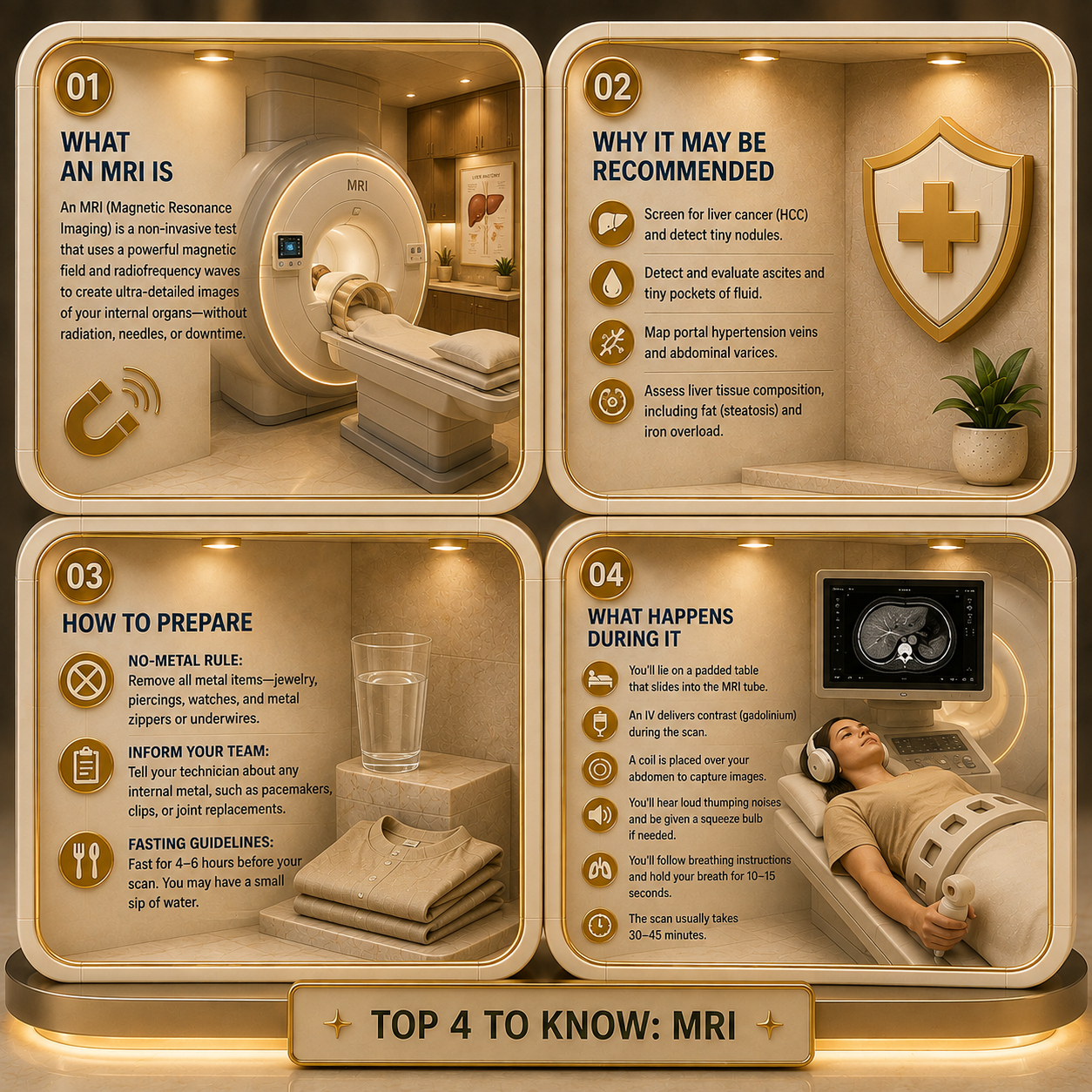
Top 4 Need to Knows
My Experience with It
By Angie from @Diagnosis_Cirrhosis on TikTok
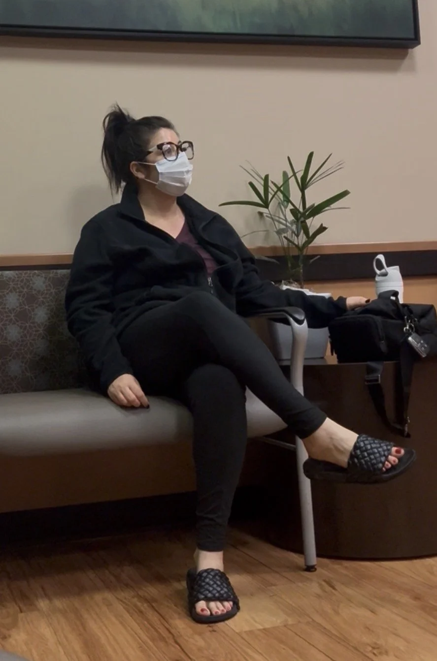
MRIs for me take place in an imaging center. My Dr. sends the order ahead of time and I schedule it. Here’s me in the waiting room last April.
Moments after hearing the word cirrhosis for the fist time, this followed:
“Okay, now you need an MRI.”
Like we were adding it to the errands list.
Order groceries.
Take out earrings.
Meet the noisy magnet.
Look at liver.
Pick up groceries.
It seemed violently casual. Abhorantly blasé. Except these days… it is.
MRI is enormous, then it’s easy. The results… those will always be stressful. The process though? Now I prefer it over food shopping to be honest.
Radiology Does Not Care About Christmas
My madden voyage through the MRI was the day after Christmas.
I had not yet learned that medical needs do not care about weekends, holidays, emotional readiness, or the fact that everyone else is still eating leftovers in pajama pants.
And the waiting room was playing Ratatouille.
Of course it was.
Because nothing says “welcome to your first liver MRI” like a rat in Paris teaching everyone that anyone can cook.
At the time, I was too scared to appreciate the comedy.
I was swollen, newly diagnosed, and carrying over 10 liters of fluid in my abdomen, a raging hemroid, and feet heavier than a bad scene in the Sopranos. .
Now I have been there enough times to know that waiting room loop.
I know what fear and familiarity look like on fellow faces.
I know where to sit without getting blasted by the AC..
I know that there’s a 70% chance a rat is trying to inspire me to make good use of my groceries.

Fear Got There First
That first time, I knew just enough to be terrified and not enough to be accurate.
I knew there was a tube.
I knew there was a needle with liquid that had special powers.
I knew I had to a forgetting earring could be catastrophic.
I knew it wouldn’t cure me like a magic trip through a medically mediated carwash express.
Yeah, I knew it probably just meant more bad news.
That was enough.
And because I did not yet have my own experience to compare it to, my imagination took the wheel, drove the car off a cliff, and somehow billed insurance on the way down.
I was so scared I asked if my husband could come back with me.
The technician looked confused.
Not rude. Just perplexed.
Like I had asked if my husband could also get into the MRI machine and hold a little umbrella over me.
Then he realized I was not mildly nervous.
I was crying hard enough to threaten the equipment.
Eventually, he said okay.
Probably because he was kind.
Probably because I looked like a liability.
Probably because nobody wants to deny a crying woman with a waterbed abdomen moral support the day after Christmas while a cartoon rat plays in the lobby.
The Scan Was Not the Monster
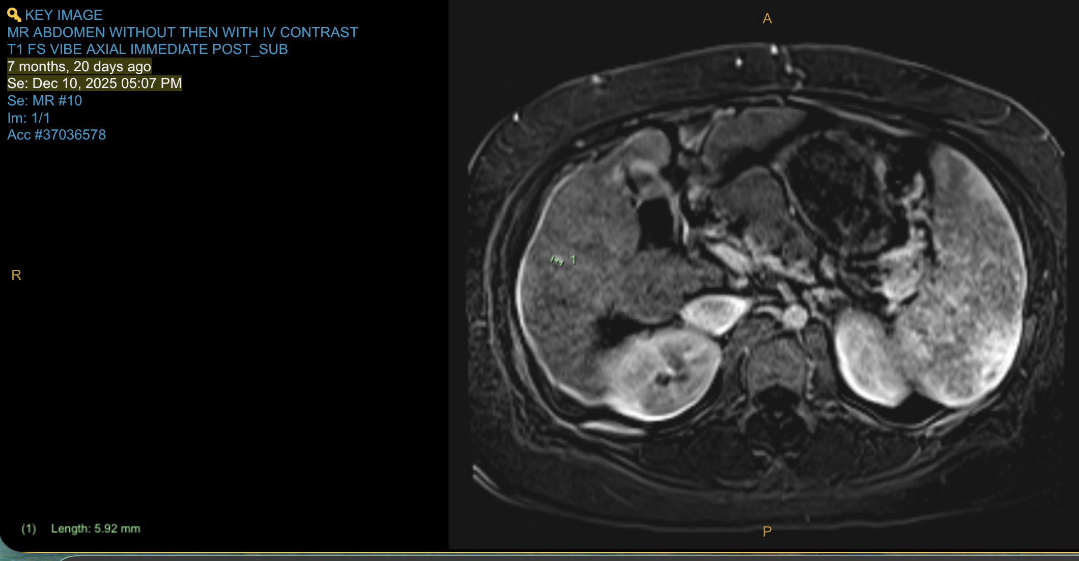
This is an image from Dec 2025. The tiny green dot is an LI-RADs 3 finding. Still TBD if it’s the thing or nothing.
Unsurprisingly, the actual MRI was nothing. To use a radiology joke, “unremarkable”.
If they use contrast, you will probably need an IV. Annoying, yes. Tragic, no.
Honestly, my imaging center places them swifter than my ER.
They position you on the table.
Theymay place something over your abdomen.
They hand you the panic button.
Then the table moves, the machine takes over, the robot talks, and you remember that this thing has a plan.
You are the cargo.
No Metal Means No Metal
The MRI is a giant magnet with strict boundaries. I think of it as an allergy to metal accessories.
No earrings.
No belly button rings.
No watches.
No underwire.
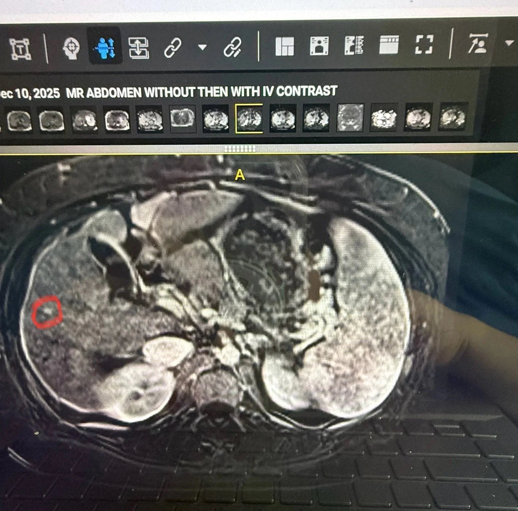
This image shows the spot, in the red circle. The LI RAD 3 finding.
No mystery zipper.
No “I think this is probably fine.”
For me, this means removing jewelry like I am preparing for a very sterile, very loud, very unsexy heist.
The Noise Is Not Music
The noise is the part nobody explained well enough to me.
The first few times, I genuinely thought radiology was playing music.
Not calming spa music.
Aggressive industrial metal. (The irony isn’t lost on me).
Bang. Clang.
Thump.
Grind. Like a chainsaw without a blade.
Like a possessed printer joined a Berlin grunge band.
And I was lying there thinking:
Wow. Radiology has a type.
It took me multiple scans to realize: Oh.
That is not music.
That is the machine.
Which somehow made it both less weird and more weird.
Nobody picked the soundtrack.
The multi-million-dollar magnet just sounds like it is trying to summon something from underneath the hospital.
Do Not Expect a Nap
People say, “Just relax, maybe you’ll nap.”
A nap?
In the magnetic jackhammer tunnel?
Absolutely not.
For liver MRIs, you usually have to participate.
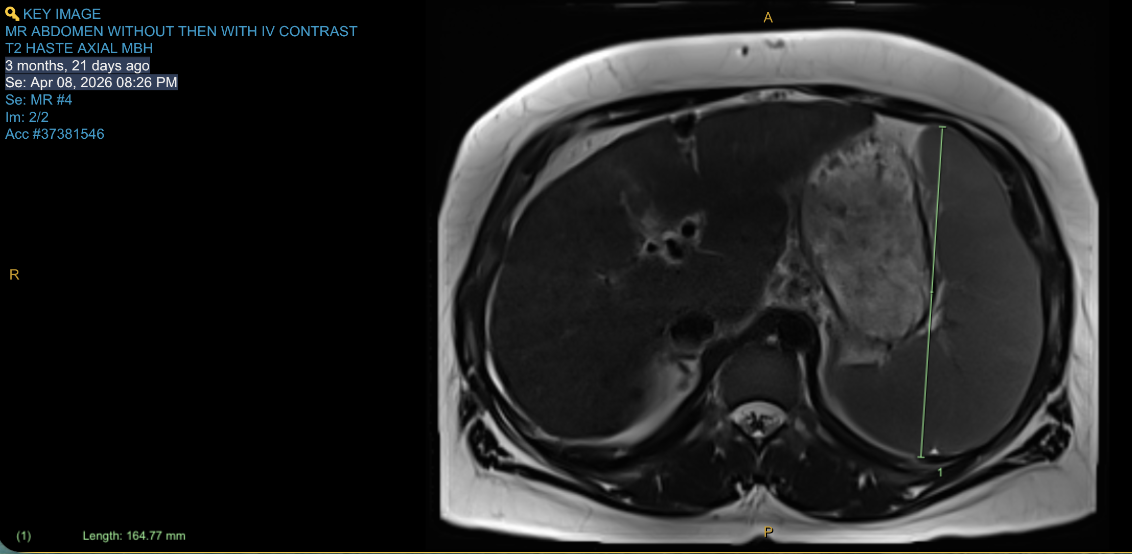
This is the quintessential view. Seems standard, but a lot going on too.
Breathe in. Hold it.
Breathe out. Hold still.
Do it again.
The breath holds matter because your liver moves when you breathe, and they need clean images.
So you are not just lying there. Very quietly. Very still.
While every cell in your body suddenly wants choreography because someone told you not to move.
My strategy is simple. I close my eyes before I go in, and I keep them closed.
I do not look around.
I do not make it a tube.
I do not let my brain measure the distance between my face and the machine.
Eyes closed.
Get through it.
And remember the panic button exists.
You are not trapped in some medieval liver tunnel.
If you truly need to stop, you can press it.
@diagnosis_cirrhosis 🩻 Why can cirrhosis be so difficult to accept? Because the damage is real—but it’s hidden. When someone breaks an arm, everyone understands what happened. There is swelling. Bruising. An X-ray. A cast. Even if you cannot see the bone itself, there is visible proof that something changed. Liver disease asks something completely different of us. Your liver sits quietly beneath your ribs. It can be scarred, permanently damaged, and struggling to function while the outside of your body may still look completely normal. That can make cirrhosis, chronic liver disease, liver damage, fibrosis, and even fatty liver disease incredibly difficult to process. You may find yourself asking: ❓What does cirrhosis actually look like? ❓Can an ultrasound show liver damage? ❓When do you need a CT scan or MRI of the liver? ❓What does a FibroScan measure? ❓Why would someone need a liver biopsy? Instead of something you can see, you’re handed blood work, imaging reports, and words like fibrosis, portal hypertension, regenerative nodules, collateral circulation, compensated cirrhosis, decompensated cirrhosis, and recompensation. Medically, those words are precise. Emotionally, they can feel almost abstract. I didn’t truly understand my diagnosis until I saw my own liver imaging. I cried. Not because the pictures were dramatic. Because for the first time, cirrhosis wasn’t just something people kept telling me I had. It had a shape. A landscape. A place. The invisible finally became visible. That image became the bridge between understanding my diagnosis intellectually and actually beginning to grieve it. 🩺 Ultrasound helps evaluate liver texture, liver contour, blood flow, portal hypertension, ascites, an enlarged spleen, and other structural changes. 🩻 CT scans provide detailed images of your liver, blood vessels, collateral veins, surrounding organs, ascites, and can help evaluate complications or liver cancer. 🧲 MRI offers even greater soft tissue detail and is often used to better characterize liver lesions, evaluate bile ducts, or clarify findings seen on ultrasound or CT. 📊 FibroScan estimates liver stiffness and liver fat, helping assess fibrosis and cirrhosis without surgery. 🔬 Liver biopsy examines a tiny sample of liver tissue under the microscope to evaluate inflammation, fibrosis, scarring, and sometimes identify the underlying cause of liver disease. Each test answers a different question. Together, they help your hepatologist monitor disease progression, screen for hepatocellular carcinoma (liver cancer), evaluate portal hypertension, detect complications, and guide treatment decisions. For some of us… They do something else, too. They help our brains catch up. Seeing your liver doesn’t change the diagnosis. But it can change your relationship with it. It gives your grief somewhere to land. Whether your cirrhosis developed from fatty liver disease (MASLD, NAFLD, MASH, or NASH) 🍩, alcohol-related liver disease 🚫🍷, hepatitis B 🦠, hepatitis C 🦠, autoimmune hepatitis 🔥, primary biliary cholangitis (PBC) 🧬, primary sclerosing cholangitis (PSC) 🧬, hemochromatosis ⚙️, Wilson disease 🪙, alpha-1 antitrypsin deficiency 🧩, medication-related liver injury 💊, vascular liver disease, or another condition, the emotional experience is often remarkably similar. You’re being asked to accept permanent liver damage you cannot touch, inspect, or watch change from one day to the next. If you’re struggling to make your diagnosis feel “real,” that doesn’t mean you’re failing at acceptance. It means your mind is trying to reconcile an invisible organ with a life that still looks visible. 👩⚕️👨⚕️🩺 Talk to your hepatologist or gastroenterologist about your liver imaging. Ask why an ultrasound, CT scan, MRI, FibroScan, or liver biopsy was recommended, what each test can and cannot show, whether your liver has changed over time, and how those findings influence treatment, surveillance, and long-term management #livehighlights #tiktoklive ♬ Dance Floor Night - Seraphine Vally
The Part That Changed
MRIs aren’t scary anymore.
The tube, the IV, the breath holds, the panic button, the possessed-printer soundtrack — all of it is fine. Normal even. “Normal”… you know what I mean.
Not pleasant. Not a spa day.
Just routine.
No metal. Except the music.
Eyes closed.
Listen to the robot.
Hold still.
Let the noisy magnet do its investigation.
Is It Still Scary?
So where’s the fear? It’s in the portal, in the report, in the phrase you do not recognize, in the tiny finding that may be nothing, may be something, may be the beginning of a hallway you did not ask to walk down.
And maybe that is the most cirrhosis thing about it.
The terrifying becomes comforting.
Because MRI turns the invisible into an image.
So yes, I still think about that first MRI.
The day after Christmas.
The waterbed abdomen.
The crying.
The cartoon rat.
But now I know this:
The machine was never the monster.
It was just loud enough to make me think it was.
Key Takeaways:
Find Out More
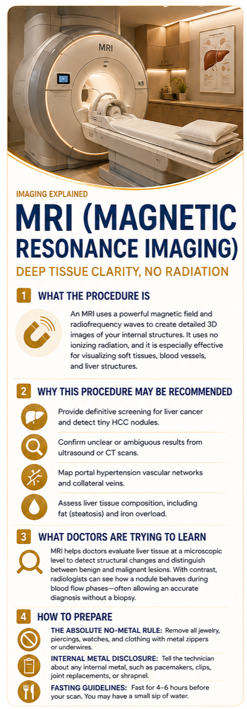
What the Procedure Is
A Magnetic Resonance Imaging, or MRI, scan is a non-invasive diagnostic technique that uses an exceptionally powerful magnetic field and radiofrequency waves to map your body’s internal structures [1]. Unlike a CT scan, an MRI uses no ionizing radiation. Instead, it temporarily realigns hydrogen atoms inside your tissues to create hyper-detailed, high-contrast three-dimensional images of your soft organs, muscles, and complex blood vessel networks [2].
Why This Procedure May Be Recommended
In advanced liver care and cirrhosis, an abdominal MRI is widely considered the gold-standard imaging tool for deep tissue evaluation. A hepatologist or transplant team will typically recommend an MRI to:
Provide definitive screening for liver cancer, as an MRI is highly capable of identifying and distinguishing tiny hepatocellular carcinoma, or HCC, nodules from benign liver tissue [3].
Confirm uncertain or ambiguous results found on a routine screening ultrasound or a rapid emergency CT scan [4].
Map out advanced portal hypertension vascular networks, tracking the exact pathways of swollen collateral veins and deep abdominal varices [3].
Assess liver tissue composition, such as measuring the precise percentage of fat accumulation, or steatosis, or iron overload in the organ [2].
What Doctors Are Trying to Learn
Physicians use an MRI to evaluate the microscopic characteristics of liver tissue and look closely at the liver’s physical architecture. While blood tests measure manufacturing function, an MRI provides the visual data needed to see the exact pattern of structural scarring.
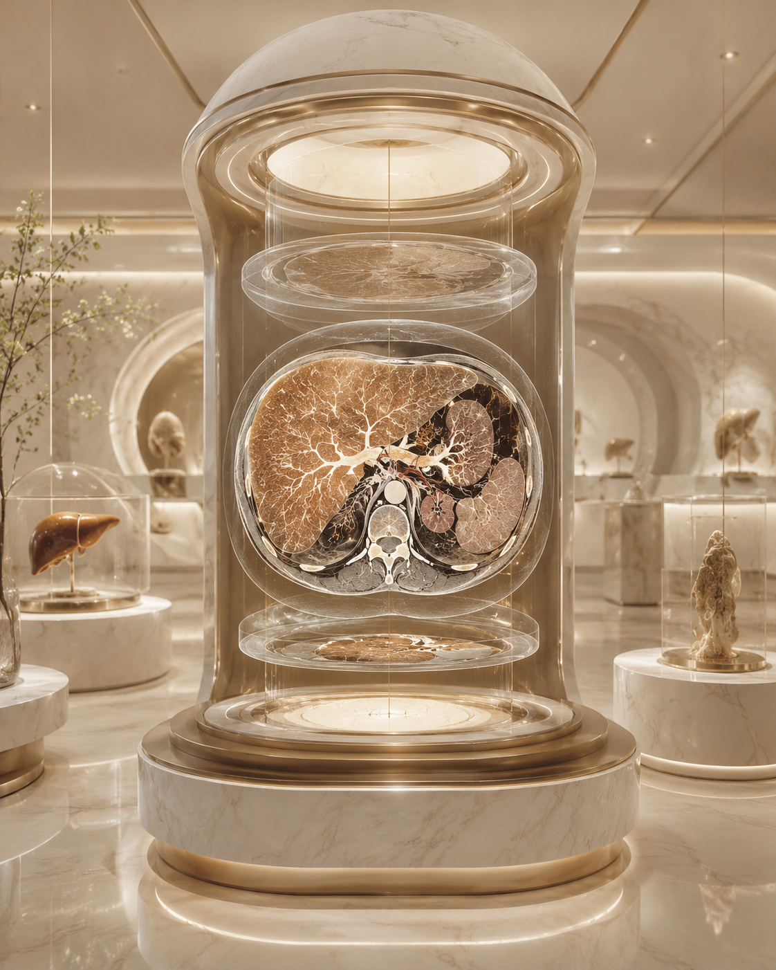
Hepatologists are trying to determine whether any structural mass or shadow is a benign liver nodule, which is common in cirrhosis, or an active, early-stage lesion that requires intervention [3]. By injecting a specialized contrast agent, usually gadolinium, radiologists can watch how a nodule “lights up” or washes out during different phases of blood flow, giving them an accurate diagnosis without needing a physical tissue needle biopsy [4].
How to Prepare
Because an MRI operates using an extremely powerful, active magnet, safety and preparation rules are strict:
The Absolute No-Metal Rule: You must remove all jewelry, earrings, watches, piercings, and clothes with metal zippers or underwires. Metal will react violently with the magnet and ruin the cross-sectional data [1].
Internal Metal Disclosure: It is vital to tell your technician if you have any internal metal, such as pacemakers, metallic clips, joint replacements, or old shrapnel [5].
Fasting Guidelines: You will typically be instructed to fast, consuming no food or drink except a small sip of water, for 4 to 6 hours before your scan to keep your gallbladder and stomach still [5].
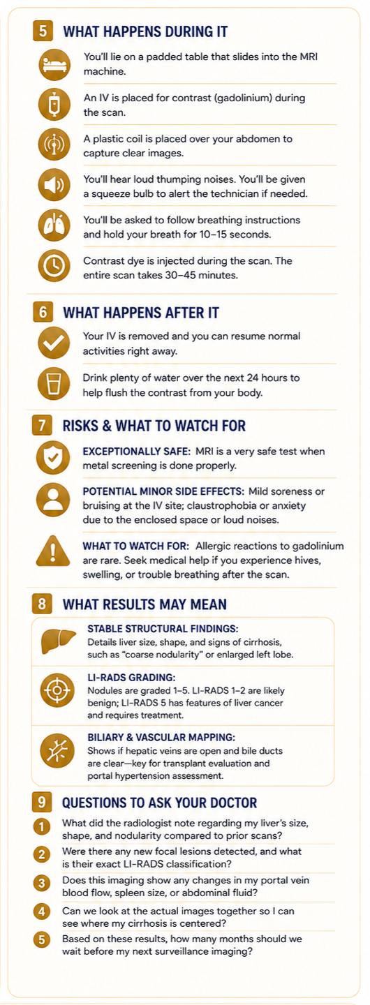
What Happens During and After
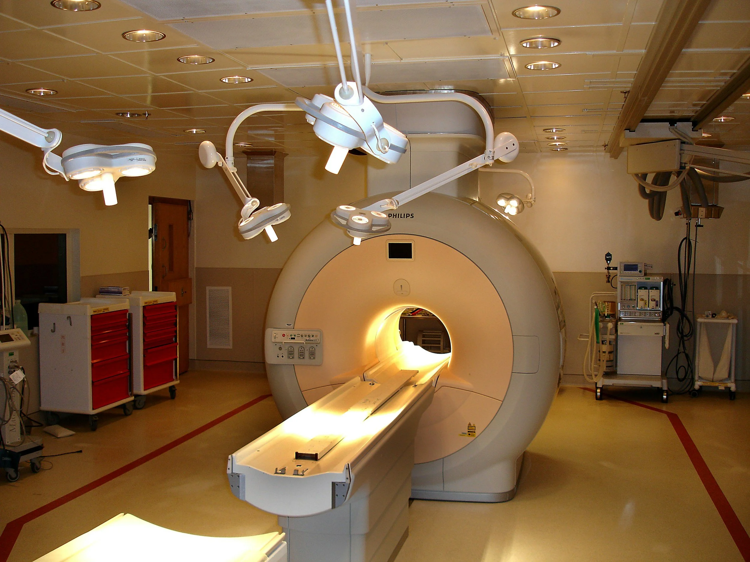
The procedure takes place inside a specialized magnetic room. A professional will place a standard intravenous, or IV, line in your arm to deliver the gadolinium contrast dye later in the scan [1]. You will lie flat on a padded motorized table that moves into a long, cylindrical tube [5].
To capture sharp liver pictures, a plastic shield containing radio antennas is placed over your abdomen, and you will be handed a physical squeeze bulb to press if you panic or need to stop [2]. Once the scan begins, the machine will emit a series of loud, rhythmic thumping and banging noises that sound like intense construction or mechanical techno music [5].
You will hear automated instructions telling you exactly when to breathe in, hold your breath for 10 to 15 seconds, and breathe out [2]. During the middle of the scan, the contrast dye is injected through your IV; unlike CT dye, it usually does not cause a heavy wave of hot flushing, though you may feel a cool sensation in your arm [1]. The entire scan takes between 30 and 45 minutes.
When it is finished, the table slides out of the cylinder, your IV line is removed, and you are free to sit up [5]. Because no sedation is required for a standard diagnostic scan, you can drive yourself home immediately, eat a normal meal, and go about your day without restrictions [2]. You will be advised to drink plenty of water over the next 24 hours to help your kidneys filter the contrast fluid out of your blood [1].
Risks and What to Watch For
An MRI scan is an exceptionally safe diagnostic test when pre-screening metal checks are performed correctly [1]. Potential minor side effects are rare and include:
Mild localized tenderness or a small bruise at the IV injection site [5].
Claustrophobia or mild anxiety caused by the loud noises or enclosed tube space [2].
Also to watch for: Allergic reactions to gadolinium contrast are incredibly rare. However, check with your clinic or seek emergency care if you experience post-scan symptoms like skin hives, swelling of the tongue, or sudden difficulty breathing [5].
What Results May Mean
A radiologist will study the thousands of raw cross-sectional images and draft a descriptive text report for your care team [5].
Stable Structural Findings
The report will such as “coarse nodularity” or an enlarged left lobe, which tells your doctor if your tissue structure is holding steady [4].
The LI-RADS Grading
If any mass or nodule is found, it will be graded on a 1-to-5 scale called the Liver Imaging Reporting and Data System, or LI-RADS. A LI-RADS 1 or 2 means the spot is a benign scar, while a LI-RADS 5 means the spot matches all visual signatures of primary liver cancer and requires an immediate treatment plan [3].
Biliary and Vascular Mapping
The report will explicitly state whether your hepatic veins are open and whether your bile ducts are clear, which are crucial metrics for transplant matching and evaluating portal hypertension [3].
Questions to Ask Your Doctor
To help build your personal tracking files and advocate for yourself at your next follow-up, consider asking your doctor these questions:
What did the radiologist note regarding my liver’s specific size, shape, and nodularity compared to my previous scans?
Were there any new focal lesions or areas of concern detected on this MRI, and if so, what is their exact LI-RADS classification?
Does this detailed imaging show any changes to my portal vein blood flow, my spleen volume, or the development of abdominal fluid?
Can we look at the actual cross-sectional images of my liver together so I can visualize where my cirrhosis is centered?
Based on these results, how many months should we wait before scheduling our next routine abdominal surveillance imaging?
Sources
[1]: Mayo Clinic: MRI Scan Overview, Magnetic Field Safety, and Gadolinium Contrast Tracking
[2]: Cleveland Clinic: Understanding Magnetic Resonance Imaging (MRI), Abdominal Soft-Tissue Cross-Sections, and Patient Care
[3]: Journal of Hepatology: Consensus Practice Guidelines on Liver Nodule Characterization, LI-RADS Classifications, and HCC Screening Protocols
[4]: The American Journal of Gastroenterology: ACG Clinical Practice Guideline: Abdominal Surveillance Imaging, Coarse Nodularity, and Management of Advanced Liver Fibrosis
[5]: National Institutes of Health (NIH) / MedlinePlus: Abdominal MRI Procedures, Preparation Protocols, Loud Machine Noises, and Patient Safety Risks
