
Ultrasound
@diagnosis_cirrhosis 📢🚨 PSA + humble plea to whoever trains ultrasound technicians out there 🚨📢 First of all — much love to the ultrasound techs of the world 🫶🩺💙 You keep the diagnostic engine of medicine running and we appreciate you more than you know. But respectfully… a few requests from the liver patient community 😅👇 1️⃣ When you say “big breath in… hold it… hold it… hold it…” could we maybe also get the memo about WHEN WE CAN BREATHE AGAIN?? 😮💨😵💫 Some of us are out here wondering if this is a medical test or a free diving competition 🏊♀️🌊 2️⃣ The wand situation… I know you have to get good images but sometimes it feels like we’re trying to dig for buried treasure through my ribs 🏴☠️🪙🔎 3️⃣ Also quick question… I personally do not know where my gallbladder is 🤷♀️ Shouldn’t you and the wand be working together to figure that out?? 🕵️♀️📡 All jokes aside — ultrasound is actually a really important tool for liver health 🩺🍃 Most people think of ultrasound as the thing used during pregnancy to see babies 👶🍼 …but it’s also one of the main ways doctors evaluate the liver. For people with cirrhosis or liver concerns, ultrasound can help doctors look for: 🔎 Signs of cirrhosis or scarring 🔎 Changes in liver size or texture 🔎 Fluid in the abdomen (ascites) 🔎 Enlarged spleen from portal hypertension 🔎 Liver nodules or tumors 🔎 Early signs of hepatocellular carcinoma (HCC) Because the liver is constantly trying to repair and regenerate, cirrhosis increases the risk of mutations that can lead to liver cancer 🔬 That’s why many people with cirrhosis get routine ultrasound screening about every 6 months 👀📅 Cirrhosis can happen for many different reasons, including: 🛡️ Autoimmune hepatitis 🍩 MASLD / NAFLD 🧪 MASH / NASH 🚫🍷 Alcohol-related liver disease 🦠 Hepatitis B 🦠 Hepatitis C 🧬 Primary biliary cholangitis (PBC) 🧬 Primary sclerosing cholangitis (PSC) ⚙️ Hemochromatosis 🪙 Wilson’s disease 🧩 Alpha-1 antitrypsin deficiency 💊 Drug-induced liver injury ❓ Cryptogenic cirrhosis …and any other causes of chronic liver damage So yes… we may joke about the wand and the breath holding 😅 But these scans are an important part of protecting liver health and catching problems early 🩺💚 And Sonja’s name and identity have been changed to protect the innocent 👀😇 #medicalstudent #radiology #LiverHealth #Ultrasound #patientcare ♬ original sound - diagnosis cirrhosis
Top 4 Need to Knows
Important Medical Disclaimer: I am Angie, a cirrhosis patient sharing my personal lifestyle, nutrition, and mindset journey. I am a data aggregator and community builder, not a licensed medical professional, doctor, or dietitian. Content on diagnosiscirrhosis.com is based solely on my personal experience living with advanced liver disease and is intended for informational and educational purposes only. It does not constitute medical advice or a substitute for professional medical care. Always follow the recommendations of your own healthcare team and consult your hepatologist or physician before changing your diet, medication, or lifestyle routine.
My Experience with It
By Angie from @Diagnosis_Cirrhosis on TikTok
The Test with Too Many Personalities
Ultrasound sounds like one test. It is not.
My whole life I thought it was for pregnancy. Happy news. Instragram.
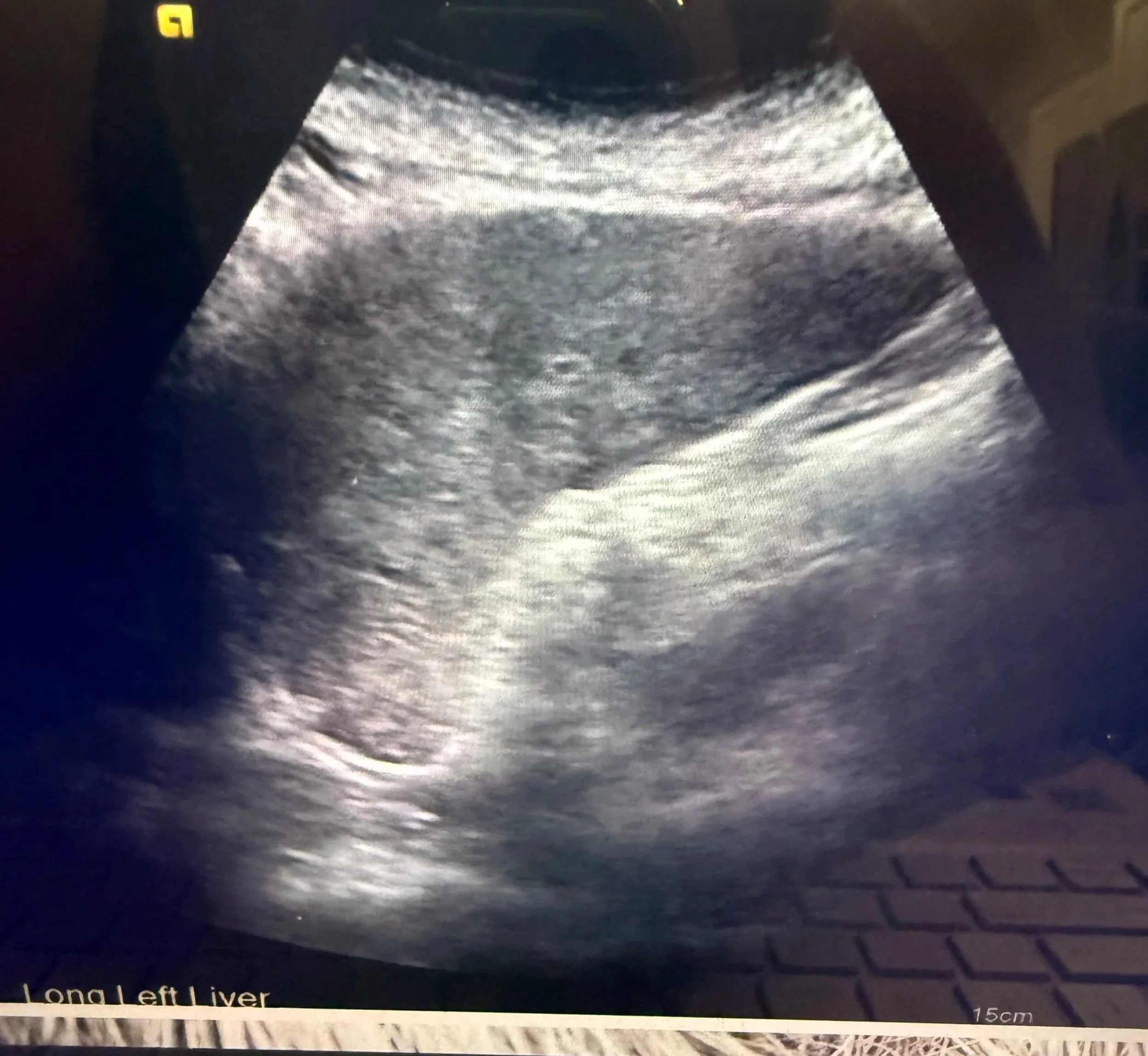
This is from the first time I saw my liver. Very memorable experience looking at the “thing” that changed my life effectively overnight.
Imagine my surprise that my first one was anything but.
Ultrasounds are a whole little universe of gel, pressure, grayscale blobs, breath-holding, awkward silence, and a technician sliding a wand around your abdomen like they are trying to find buried treasure in a haunted field.
As if the quick bedside scan checking for fluid or a baby is the same experience as the full abdominal ultrasound where they are inspecting your liver, spleen, bile ducts, vessels, texture, shadows, and whatever suspicious little grayscale nonsense decides to make an appearance that day.
Same tool. Very different mission.
Because even in cirrhosis, ultrasound can show up in completely different ways depending on what question the doctor is trying to answer.
Sometimes the question is:
Do you have ascites right now?
Sometimes it is:
Where is the safest pocket of fluid before a paracentesis?
Sometimes it is:
Are we seeing anything concerning for liver cancer surveillance?
Sometimes it is:
Does this look stable, or do we need a closer look with CT or MRI?
It is like the Swiss Army knife of liver imaging.
Not glamorous. Not dramatic.
Not usually the test people write orchestral music about.
But constantly useful.
The Fluid Check
In the hospital, ultrasound is often used to check for ascites — fluid that can build up in the abdomen with cirrhosis.
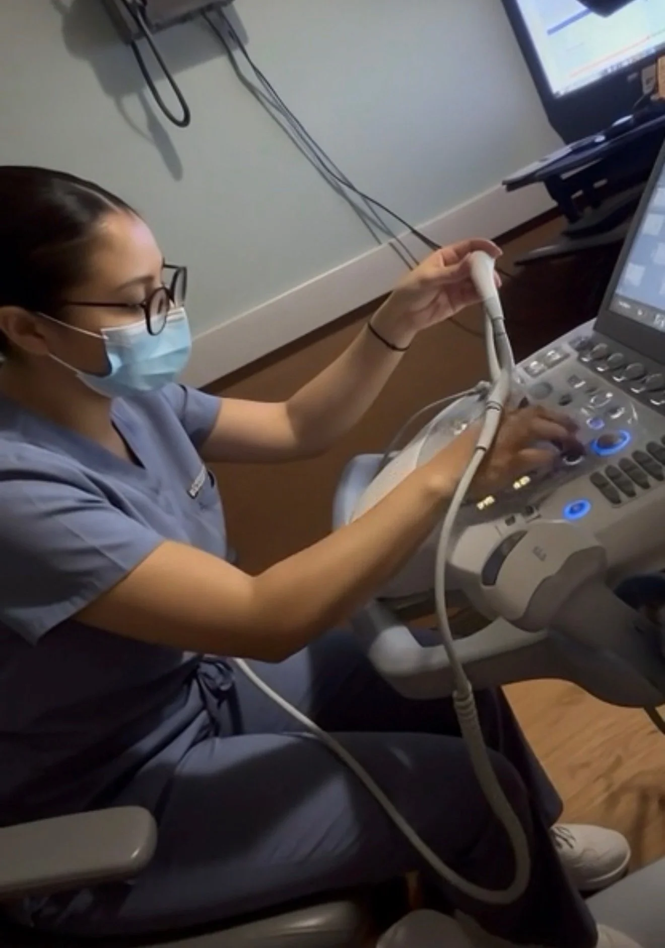
The technician at my last ultrasound was kind enough to show me the machine nd explain a few things before getting started.
If your belly feels tight, hard, swollen, or suspiciously wrong, ultrasound can help confirm whether fluid is actually there.
It is also commonly used before a paracentesis.
If someone is going to place a needle into your abdomen and drain fluid, they need to know where the fluid is and where it is safest to go.
Which I appreciate. Very much.
Let’s not freestyle with the abdominal needle.
The Formal Liver Ultrasounds
A formal abdominal ultrasound is different from a quick fluid check.
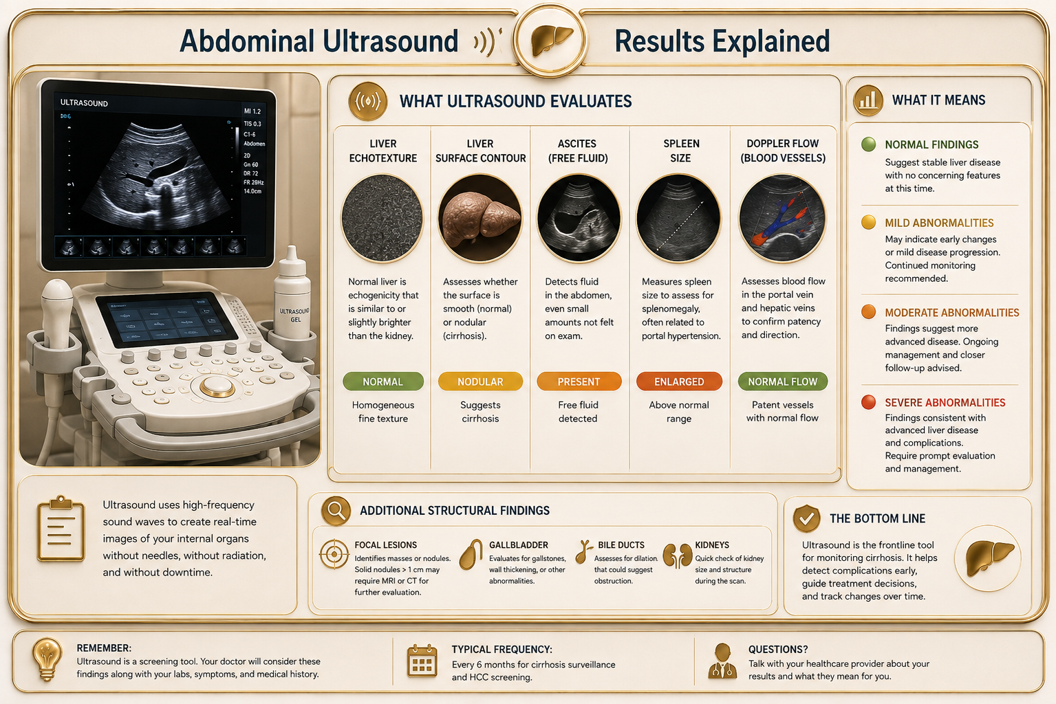
That scan is usually looking more broadly at the liver and surrounding structures: liver texture, ascites, spleen size, bile ducts, blood flow, and anything that may need a closer look.
For many cirrhosis patients,ultrasound is also part of routine HCC surveillance, which means liver cancer screening.
That does not mean they think you have cancer every time they order it.
It means cirrhosis increases the risk, so the liver has to be watched.
Jump to Key Takeaways
Sometimes this scan also includes Doppler, which is the part that looks at blood flow.
That matters in cirrhosis because portal hypertension is basically a traffic disaster in vascular form. Doppler can help doctors see how blood is moving through important vessels and whether the flow looks normal, reversed, blocked, or rerouted.
So if you hear strange whooshing sounds during the scan, that may be Doppler.
Very casual. Very submarine.
Very “why is my abdomen making weather noises?”
But it is useful information.
For me, Doppler made portal hypertension feel less abstract. It was not just a phrase anymore. It was blood flow, detours, pressure, and old pathways reopening because the normal route through my liver had become a disaster zone.
That distinction helped me. Same wand.
Different mission.
What It Actually Feels Like
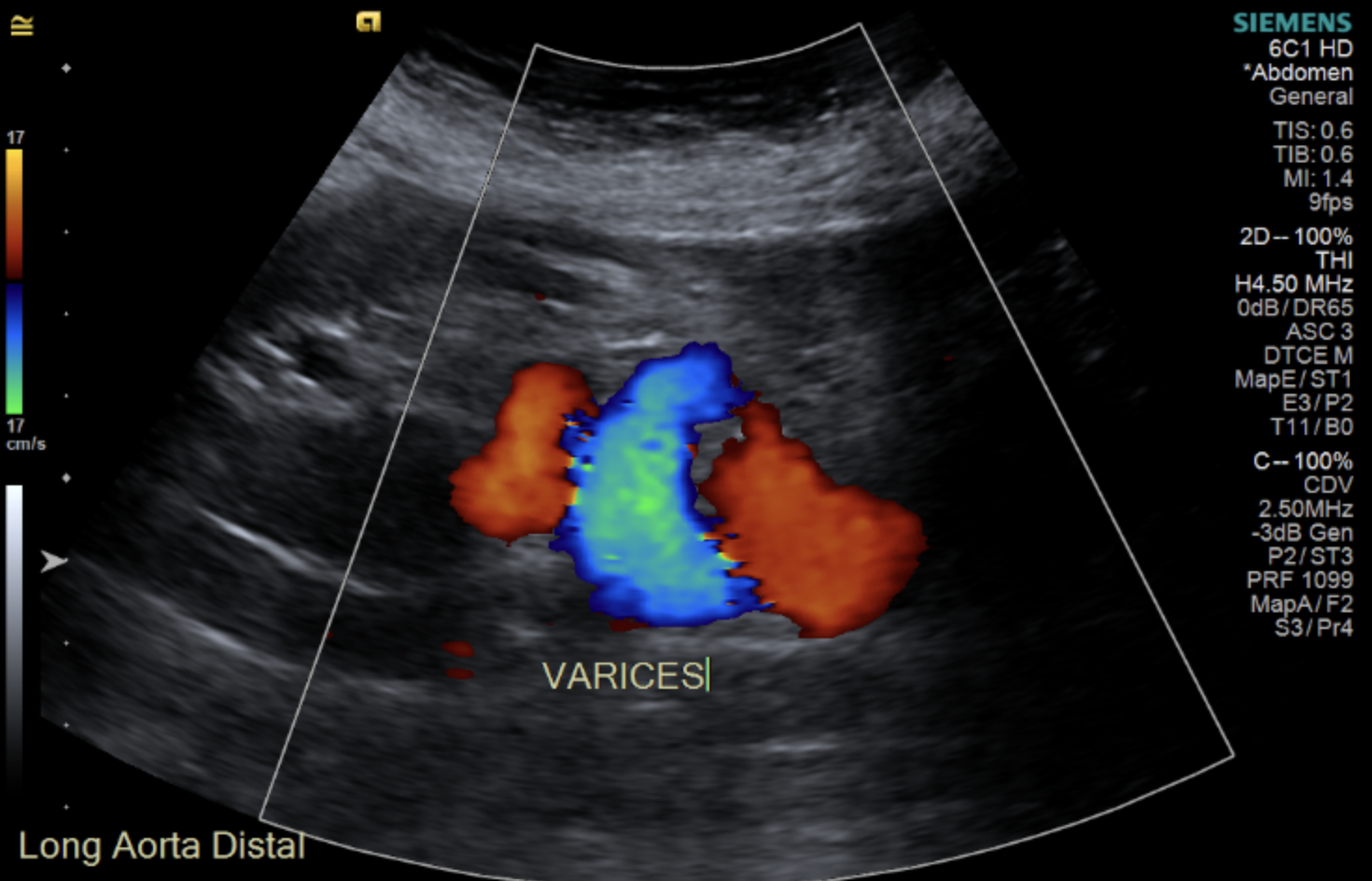
Doppler ultrasounds are known for a whooshing sound!
Ultrasound is usually described as painless.
Technically, yes.
No needles.
No contrast dye.
No giant MRI tube.
No CT warmth that makes you briefly question your drive home.
But painless does not always mean comfortable.
The tech may press under your ribs, along your side, or into areas that already feel tender or bloated.
There have been times I was lying there trying to be mature and medically cooperative while thinking:
That is my organ.
That is still my organ.
Why are we trying to push it into the table?
And then there is the breathing.
Breathe in.
Hold it.
Let it out.
Hold again.
Repeat many, many times.
Sometimes the tech is focused on the screen and you are still lying there holding your breath like a Victorian child trying to win a fainting contest.
@diagnosis_cirrhosis 🩻 Why can cirrhosis be so difficult to accept? Because the damage is real—but it’s hidden. When someone breaks an arm, everyone understands what happened. There is swelling. Bruising. An X-ray. A cast. Even if you cannot see the bone itself, there is visible proof that something changed. Liver disease asks something completely different of us. Your liver sits quietly beneath your ribs. It can be scarred, permanently damaged, and struggling to function while the outside of your body may still look completely normal. That can make cirrhosis, chronic liver disease, liver damage, fibrosis, and even fatty liver disease incredibly difficult to process. You may find yourself asking: ❓What does cirrhosis actually look like? ❓Can an ultrasound show liver damage? ❓When do you need a CT scan or MRI of the liver? ❓What does a FibroScan measure? ❓Why would someone need a liver biopsy? Instead of something you can see, you’re handed blood work, imaging reports, and words like fibrosis, portal hypertension, regenerative nodules, collateral circulation, compensated cirrhosis, decompensated cirrhosis, and recompensation. Medically, those words are precise. Emotionally, they can feel almost abstract. I didn’t truly understand my diagnosis until I saw my own liver imaging. I cried. Not because the pictures were dramatic. Because for the first time, cirrhosis wasn’t just something people kept telling me I had. It had a shape. A landscape. A place. The invisible finally became visible. That image became the bridge between understanding my diagnosis intellectually and actually beginning to grieve it. 🩺 Ultrasound helps evaluate liver texture, liver contour, blood flow, portal hypertension, ascites, an enlarged spleen, and other structural changes. 🩻 CT scans provide detailed images of your liver, blood vessels, collateral veins, surrounding organs, ascites, and can help evaluate complications or liver cancer. 🧲 MRI offers even greater soft tissue detail and is often used to better characterize liver lesions, evaluate bile ducts, or clarify findings seen on ultrasound or CT. 📊 FibroScan estimates liver stiffness and liver fat, helping assess fibrosis and cirrhosis without surgery. 🔬 Liver biopsy examines a tiny sample of liver tissue under the microscope to evaluate inflammation, fibrosis, scarring, and sometimes identify the underlying cause of liver disease. Each test answers a different question. Together, they help your hepatologist monitor disease progression, screen for hepatocellular carcinoma (liver cancer), evaluate portal hypertension, detect complications, and guide treatment decisions. For some of us… They do something else, too. They help our brains catch up. Seeing your liver doesn’t change the diagnosis. But it can change your relationship with it. It gives your grief somewhere to land. Whether your cirrhosis developed from fatty liver disease (MASLD, NAFLD, MASH, or NASH) 🍩, alcohol-related liver disease 🚫🍷, hepatitis B 🦠, hepatitis C 🦠, autoimmune hepatitis 🔥, primary biliary cholangitis (PBC) 🧬, primary sclerosing cholangitis (PSC) 🧬, hemochromatosis ⚙️, Wilson disease 🪙, alpha-1 antitrypsin deficiency 🧩, medication-related liver injury 💊, vascular liver disease, or another condition, the emotional experience is often remarkably similar. You’re being asked to accept permanent liver damage you cannot touch, inspect, or watch change from one day to the next. If you’re struggling to make your diagnosis feel “real,” that doesn’t mean you’re failing at acceptance. It means your mind is trying to reconcile an invisible organ with a life that still looks visible. 👩⚕️👨⚕️🩺 Talk to your hepatologist or gastroenterologist about your liver imaging. Ask why an ultrasound, CT scan, MRI, FibroScan, or liver biopsy was recommended, what each test can and cannot show, whether your liver has changed over time, and how those findings influence treatment, surveillance, and long-term management #livehighlights #tiktoklive ♬ Dance Floor Night - Seraphine Vally
You are allowed to say something.
Ask for a break.
Say the pressure is too much.
Say, “I need to breathe.”
You are not being difficult.
You are the body on the table.
The body gets a vote.
The Magic Camera Thing
Ultrasound has also given me some strange little cirrhosis comedy moments.
One time, a tech asked me if I still had my gallbladder.
And I remember thinking:
Aren’t you the one with the magic camera thing?
I brought the abdomen.
You brought the wand.
Surely one of us is better equipped to solve this mystery.
Another time, a tech saw my recanalized umbilical vein and got very excited.
Which is alarming when you are the person attached to the finding.
A recanalized umbilical vein is related to portal hypertension. It can happen when blood flow through the liver is under high pressure and the body reopens older collateral pathways.
But when someone says “umbilical” with enthusiasm while scanning your cirrhotic abdomen, your brain does not calmly produce vascular anatomy.
Your brain goes:
Umbilical?
As in cord?
As in baby + me?
As in do I need to call someone?
Keep in mind my only reference for a long time was babies. Startling is an understatement.
That is why ultrasound can be strange.
It is routine, but it can still introduce you to words you were not emotionally prepared to Google.
Routine Does Not Mean Empty
Because ultrasound is common, it is easy to underestimate.
Don’t.
It may not feel dramatic.
It may not look dramatic.
But a lot can happen in that dim little room with the gel, the wand, and the grayscale screen.
And it’s ok if it feels like a lot sometimes.
For me, ultrasound is the test that taught me that non-invasive does not mean emotionally neutral.
Sometimes the quietest test in the room is still carrying very serious information.
Key Takeaways:
Find Out More
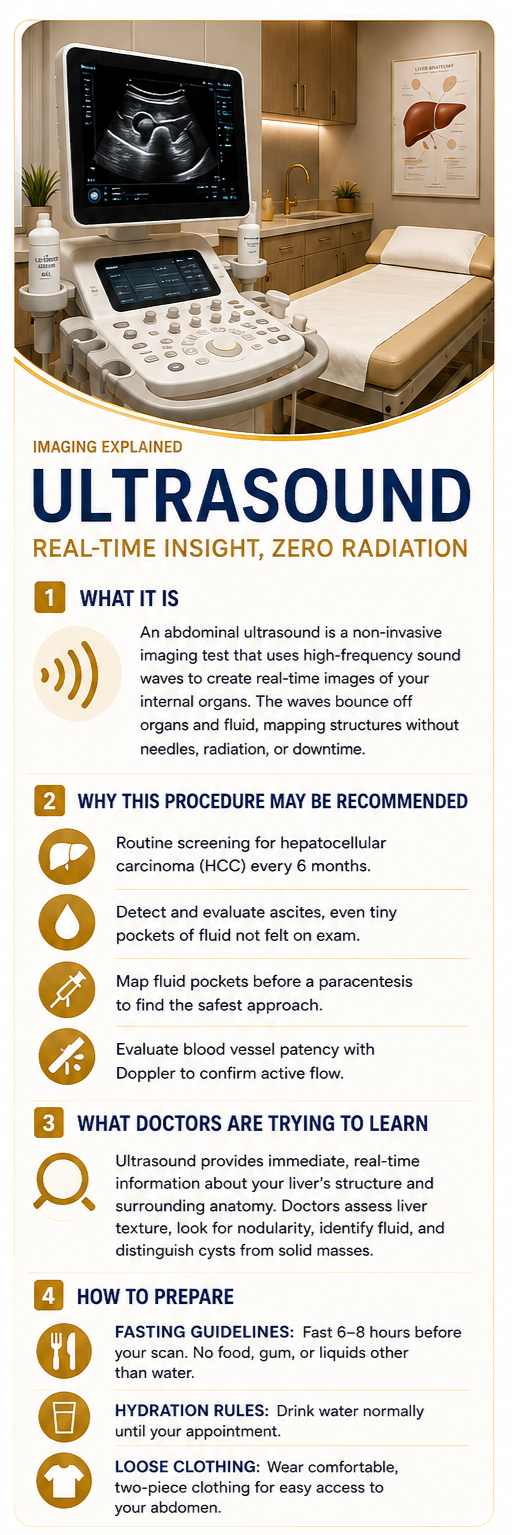
What the Procedure Is
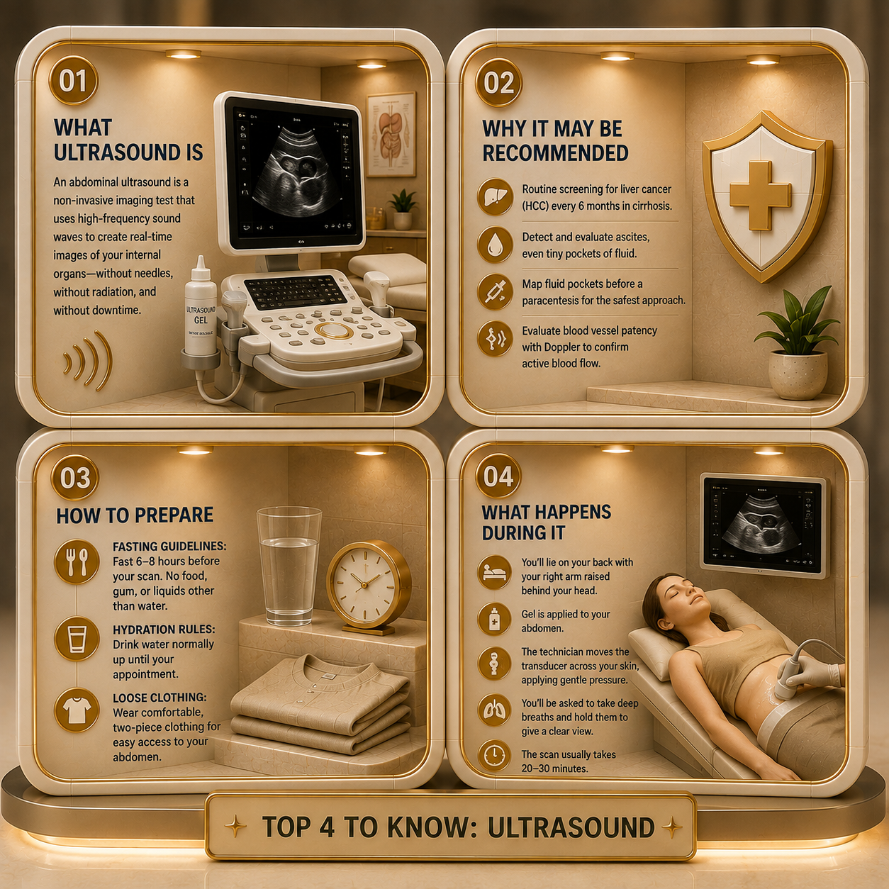
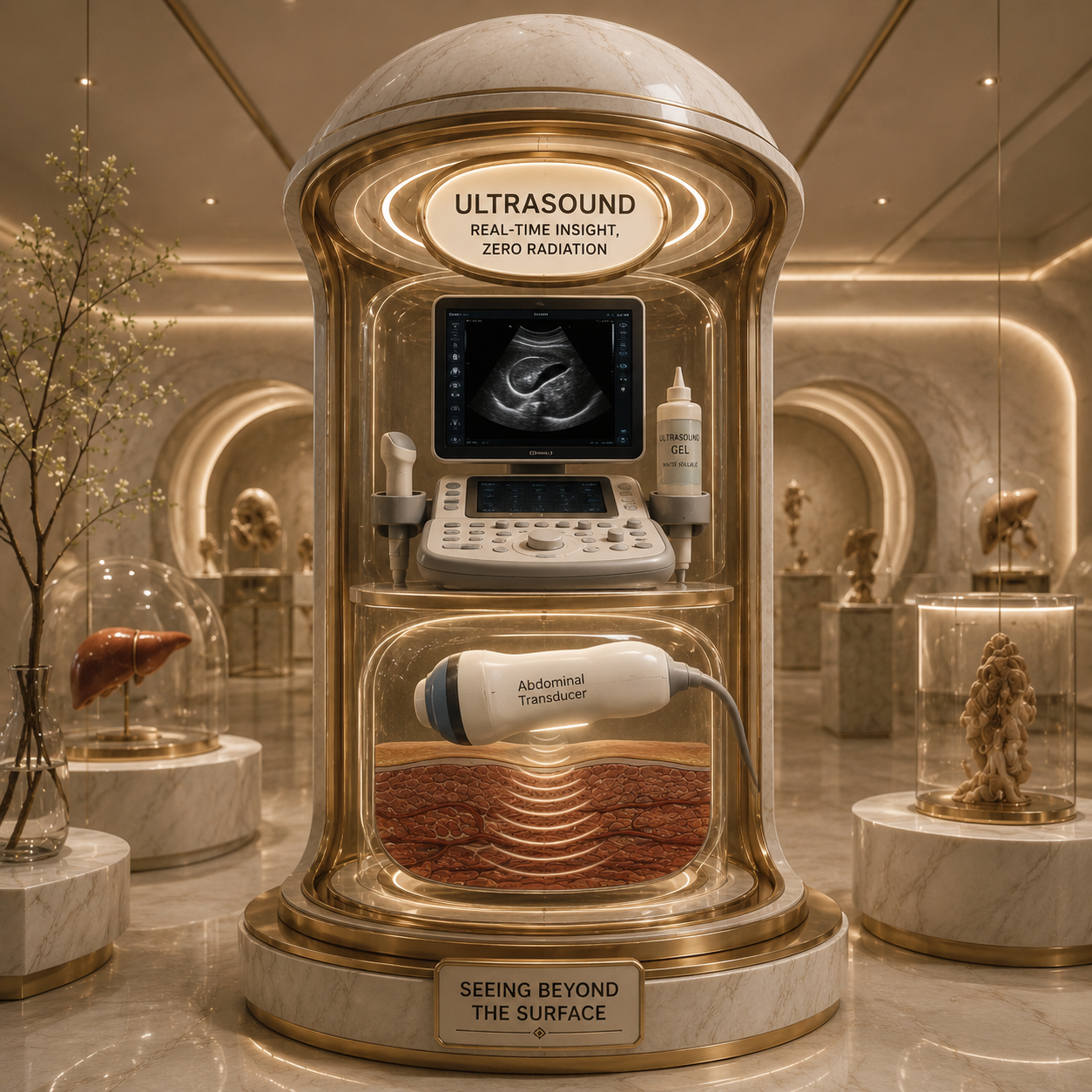
An abdominal ultrasound is a non-invasive diagnostic imaging test that utilizes high-frequency sound waves to create real-time visual images of your internal organs [1]. A hand-held device called a transducer is pressed against the skin; it emits sound waves that travel into the body, bounce off solid organs and fluid collections, and return to a computer to map out physical structures without using any ionizing radiation [2].
Why This Procedure May Be Recommended
In the clinical care of cirrhosis, the abdominal ultrasound is the single most widely used frontline monitoring tool. A medical provider will recommend an ultrasound to:
Conduct routine screening for hepatocellular carcinoma, or HCC, as international liver consensus guidelines mandate a screening ultrasound every 6 months for all cirrhosis patients [3].
Detect and evaluate the presence of ascites, identifying even tiny pockets of free fluid in the peritoneal cavity that cannot be felt during a physical examination [4].
Perform pre-procedural mapping directly before an abdominal paracentesis to mark the safest pockets of fluid and avoid accidental needle damage to internal organs [5].
Evaluate the patency of blood vessels using a specialized Doppler ultrasound to ensure that blood is actively moving through the hepatic veins and portal vein system [3].
What Doctors Are Trying to Learn
@diagnosis_cirrhosis 🎥 One more clip from the live I want to share — the realities of cirrhosis and its relationship to liver cancer, specifically HCC (hepatocellular carcinoma). ⚠️📚 This comes up a lot in the cirrhosis community, so it’s important to talk about it honestly and clearly. 💬🧠📖 When someone has cirrhosis, the risk of developing liver cancer (HCC) is higher than in the general population. That’s why people with cirrhosis are usually monitored regularly. 👀👀👀 Most hepatologists recommend screening every 6 months using imaging like ultrasound, CT, or MRI — sometimes along with blood tests like AFP — so that if something develops, it can be caught early. 🏥🩻🧪📊 A few quick facts: 📊 About 80–90% of liver cancer cases occur in people who already have cirrhosis. 📊 Research suggests that around 1 in 6 people with cirrhosis will develop some form of HCC over their lifetime. 📊 One reason this risk exists is that the liver is constantly trying to repair and regenerate damaged tissue. When cells are repeatedly dividing and repairing, mutations are more likely to occur, which can sometimes lead to cancer. 🔬🧬🧫 📊 Because of regular surveillance and screening, many cases are caught earlier, when treatment options and outcomes can be much better. 👀👀📈 That’s why monitoring matters. It’s not about expecting the worst — it’s about watching carefully so problems can be caught early if they happen. 👀👀👀 Just like cirrhosis itself, HCC can develop from many underlying liver diseases, including: Autoimmune hepatitis 🔥 MASLD / NAFLD 🍩 MASH / NASH 🧪 Alcohol-related liver disease 🚫🍷 Hepatitis B 🦠 Hepatitis C 🦠 Primary biliary cholangitis 🧬 Primary sclerosing cholangitis 🧬 Hemochromatosis ⚙️ Wilson’s disease 🪙 Alpha-1 antitrypsin deficiency 🧩 Drug-induced liver injury 💊 Fatty liver disease 🍔 Metabolic or genetic conditions 🧬 Biliary diseases 🧪 Cardiac / congestive liver disease 🏥 …or any other causes. For many people living with cirrhosis, regular monitoring becomes part of life — labs, imaging, and check-ins with hepatology teams. 🏥🧪📊👀 And while the word “cancer” is scary, surveillance and modern treatments have changed outcomes dramatically over time. 📈🔬🧬 That’s why conversations like the one we had on the live matter so much — sharing information, asking questions, and helping people understand what to watch for and why these screenings exist. 💬📚🧠👀 Knowledge doesn’t remove the uncertainty, but it helps people navigate it with better tools and better support. 🙌📚 #tiktoklive #livehighlight #cancerawareness #liverhealth #autoimmunedisease ♬ Knowledge - Vin Music
Physicians use an ultrasound to gather immediate, real-time data about the structural dimensions of your liver and surrounding abdominal anatomy [2]. While blood panels give insight into synthetic manufacturing, the ultrasound reveals the physical texture of the organ [4].
Hepatologists are trying to determine if the liver borders appear smooth or demonstrate the rough, coarse nodularity typical of advanced scarring [4]. Furthermore, because sound waves travel perfectly through liquid but bounce sharply off solid objects, doctors use ultrasound to differentiate between a benign, fluid-filled cyst and a suspicious, solid tissue mass that requires a follow-up MRI or CT scan [3].
How to Prepare
While an ultrasound is entirely non-invasive, following the correct preparation rules prevents gas and food from blocking the technician’s view:
Fasting Guidelines: You will typically be instructed to fast, consuming no food, gum, or liquids other than water, for 6 to 8 hours before an upper abdominal ultrasound to prevent bowel gas from shadowing your liver and pancreas [5].
Hydration Rules: Drink water normally up until your appointment; staying hydrated can make it easier for the technician to capture clean, sharp images [1].
Loose Clothing: Wear comfortable, two-piece clothing so that your abdomen can be easily accessed without needing to change into a full hospital gown [2].
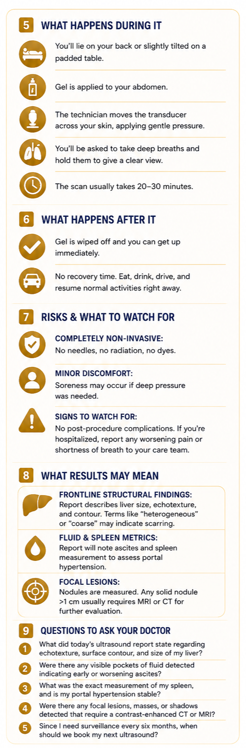
What Happens During It
The procedure takes place in a dimmed ultrasound room and is completely free of needles [5].
You will lie flat or slightly tilted on a padded examination table [5].
The sonographer will apply a generous amount of clear, water-soluble conductive gel directly onto your bare abdomen, which might feel slightly cool at first [1].
As the technician firmly slides and angles the plastic transducer wand across your skin, they will often apply deep, localized pressure to push past bowel gas, particularly when angling under your rib cage to view your kidneys or spleen [2].
Throughout the scan, the technician will give you frequent instructions to take a deep breath and hold it for several seconds; this pushes the liver downward past your ribs, giving the machine a perfectly clear window [2].
The entire process is usually completed in 20 to 30 minutes.
What Happens After It
Once the imaging is finished, the technician will hand you a paper towel or cloth to wipe the remaining gel off your skin [5]. Because no contrast dyes or sedation are used, there is absolutely no recovery period; you can leave the clinic immediately, drive yourself home, eat a normal meal, and resume all of your regular daily tasks [1].
Risks and What to Watch For
An abdominal ultrasound carries an exceptionally clean safety profile with zero clinical risks [1].
No Radiation or Dyes: Because the system operates entirely on harmless acoustic sound waves, there is no exposure to X-ray radiation and no risk of kidney stress from contrast dyes [2].
Localized Discomfort: The only real risk is minor physical soreness or temporary tenderness if the sonographer had to press heavily against a sore abdomen or an enlarged spleen [5].
Signs to watch for: There are no post-procedure complications to monitor. However, if you are getting an ultrasound as part of an active stay in the hospital, always report any worsening baseline abdominal pain or severe shortness of breath to your nursing staff [4].
What Results May Mean
A specialized radiologist will review the frozen ultrasound frames and submit a typed diagnostic summary to your hepatologist [5].
Frontline Structural Findings
The report will describe the physical dimensions of your liver, often using terms like “heterogeneous,” “echogenic,” or “coarse echotexture,” which are standard ultrasound phrases indicating that healthy liver tissue has been replaced by cirrhotic scar tissue [4].
Fluid and Spleen Metrics
The radiologist will state whether an “anechoic,” or fluid-filled, space is visible in the abdomen, confirming active ascites, and will measure your spleen in centimeters to check for splenomegaly [4].
Focal Lesions
If a new shadow or nodule is detected, it will be documented with exact measurements. Because ultrasound is a screening tool rather than a definitive diagnostic tool, any new solid nodule greater than 1 centimeter will typically trigger an immediate order for a high-resolution contrast MRI or CT scan to formally rule out liver cancer [3].
Questions to Ask Your Doctor
To help track your routine surveillance files and advocate for your care at your next clinical follow-up, consider asking your doctor these questions:
What did today’s ultrasound report state regarding the overall echotexture, surface contour, and size of my liver?
Were there any visible pockets of fluid detected on this scan that would indicate early or worsening ascites?
What was the exact measurement of my spleen, and does it show that my portal hypertension is holding stable compared to last time?
Were there any focal lesions, masses, or shadows detected that require us to step up to a contrast-enhanced CT or MRI scan?
Since my screening schedule requires surveillance every six months, when should we officially open the window to book my next routine ultrasound?
Sources
[1] Review the core clinical indications, imaging expectations, and baseline preparation rules via the Mayo Clinic Abdominal Ultrasound Guide.
[2] Study the underlying purpose, diagnostic reach, and typical durations of abdomen scans on the Cleveland Clinic Abdominal Ultrasound Portal.
[3] Read about screening standards, biannual timeline modifications, and early-stage surveillance recommendations at the Gastroenterology & Endoscopy News Summary of EASL/AASLD Practices.
[4] Access official guidelines regarding parenchymal monitoring, ascites identification, and liver texture assessments at the National Institutes of Health PMC Non-Invasive Ultrasound Assessment Review.
[5] View fundamental guidelines on general imaging preparation, transducer types, and air-pocket reduction methods through the Mayo Clinic General Ultrasound Procedures Guide.
